


The crystal structure of the N-terminal domain of MdmX in complex with p53p peptide fragment
Cheng, X.Y., Zhang, B.L., Li, Z.C., Kuang, Z.K., Su, Z.D.To be published.
Experimental Data Snapshot
Starting Model: experimental
View more details
Entity ID: 1 | |||||
|---|---|---|---|---|---|
| Molecule | Chains | Sequence Length | Organism | Details | Image |
| Cellular tumor antigen p53,Protein Mdm4 | 130 | Homo sapiens | Mutation(s): 0 | 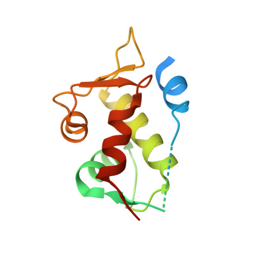 | |
UniProt & NIH Common Fund Data Resources | |||||
PHAROS: O15151 GTEx: ENSG00000198625 | |||||
PHAROS: P04637 GTEx: ENSG00000141510 | |||||
Entity Groups | |||||
| Sequence Clusters | 30% Identity50% Identity70% Identity90% Identity95% Identity100% Identity | ||||
| UniProt Groups | O15151P04637 | ||||
Sequence AnnotationsExpand | |||||
Reference Sequence | |||||
| Ligands 2 Unique | |||||
|---|---|---|---|---|---|
| ID | Chains | Name / Formula / InChI Key | 2D Diagram | 3D Interactions | |
| O4B Download:Ideal Coordinates CCD File | B [auth A], C [auth A] | 1,4,7,10,13,16-HEXAOXACYCLOOCTADECANE C12 H24 O6 XEZNGIUYQVAUSS-UHFFFAOYSA-N |  | ||
| MG Download:Ideal Coordinates CCD File | D [auth A], E [auth A] | MAGNESIUM ION Mg JLVVSXFLKOJNIY-UHFFFAOYSA-N |  | ||
| Length ( Å ) | Angle ( ˚ ) |
|---|---|
| a = 65.097 | α = 90 |
| b = 65.097 | β = 90 |
| c = 94.977 | γ = 90 |
| Software Name | Purpose |
|---|---|
| REFMAC | refinement |
| HKL-2000 | data collection |
| autoPROC | data processing |
| XDS | data reduction |
| XSCALE | data scaling |
| PDB_EXTRACT | data extraction |


