Structural analysis of the activation and DNA interactions of the response regulator VbrR from Vibrio parahaemolyticus.
Cho, S.Y., Yoon, S.I.(2021) Biochem Biophys Res Commun 555: 102-108
- PubMed: 33813268 Search on PubMed
- DOI: https://doi.org/10.1016/j.bbrc.2021.03.114
- Primary Citation Related Structures:
7E90, 7E92 - PubMed Abstract:
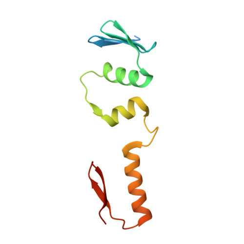
VbrK and VbrR from the gastroenteritis-causing Vibrio parahaemolyticus are a histidine kinase and response regulator, respectively, that constitute a two-component regulatory system. VbrK responds to β-lactam antibiotics or nitrate and activates VbrR via phosphorylation. Consequently, VbrR transcriptionally regulates the expression of β-lactamase and ExsC and contributes to the survival or virulence of V. parahaemolyticus. Due to the unavailability of the VbrR structure, it remains unclear how VbrR is activated via its N-terminal receiver domain (RD) and recognizes dsDNA via its C-terminal DNA-binding domain (DBD). To reveal the mechanism underlying VbrR-mediated activation, we generated the phosphomimetic protein (VbrR RD-D51E ) of the VbrR RD by replacing the D51 residue at the phosphorylation site with glutamate. VbrR RD-D51E exhibits a β 7 α 5 structure rather than the typical β 5 α 5 structure because it contains a unique two-stranded β-sheet. The VbrR RD-D51E structure represents an active state in which the D51E residue interacts with the T78 residue. As a result, the Y97 residue adopts an inward conformation, allowing VbrR RD-D51E to dimerize using the α4-β5-α5 face. These activation events are facilitated by a VbrR-specific residue, R52. Further structural study demonstrated that the VbrR DBD adopts a β-strand-decorated three-helix structure. Based on a comparative structural study, we propose that VbrR recognizes dsDNA by inserting the α8 helix into the major groove of dsDNA and interacting with the minor groove of dsDNA via the β11-β12 region. Our findings will provide a new avenue for development of new antibacterial drugs for treating V. parahaemolyticus infections.
- Division of Biomedical Convergence, College of Biomedical Science, Kangwon National University, Chuncheon, 24341, Republic of Korea.
Organizational Affiliation: