7E5V | pdb_00007e5v

Crystal structure of Phm7 in complex with inhibitor


Experimental Data Snapshot

  • Method: X-RAY DIFFRACTION
  • Resolution: 1.61 Å
  • R-Value Free: 
    0.211 (Depositor), 0.216 (DCC) 
  • R-Value Work: 
    0.183 (Depositor), 0.192 (DCC) 

Starting Model: experimental
View more details

wwPDB Validation 3D Report Full Report

Validation slider image for 7E5V

Ligand Structure Quality Assessment 


This is version 1.2 of the entry. See complete history

Literature

Molecular Basis for Two Stereoselective Diels-Alderases that Produce Decalin Skeletons*.

Fujiyama, K.Kato, N.Re, S.Kinugasa, K.Watanabe, K.Takita, R.Nogawa, T.Hino, T.Osada, H.Sugita, Y.Takahashi, S.Nagano, S.

(2021) Angew Chem Int Ed Engl 60: 22401-22410

  • DOI: https://doi.org/10.1002/anie.202106186
  • Primary Citation Related Structures: 
    7E5T, 7E5U, 7E5V

  • PubMed Abstract: 

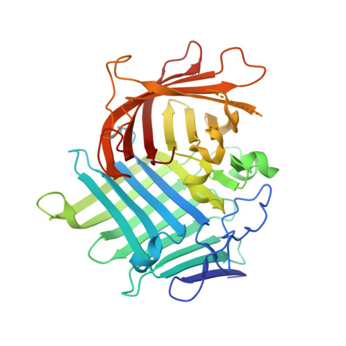
    Enzymes catalyzing [4+2] cycloaddition have attracted increasing attention because of their key roles in natural product biosynthesis. Here, we solved the X-ray crystal structures of a pair of decalin synthases, Fsa2 and Phm7, that catalyze intramolecular [4+2] cycloadditions to form enantiomeric decalin scaffolds during biosynthesis of the HIV-1 integrase inhibitor equisetin and its stereochemical opposite, phomasetin. Computational modeling, using molecular dynamics simulations as well as quantum chemical calculations, demonstrates that the reactions proceed through synergetic conformational constraints assuring transition state-like substrates folds and their stabilization by specific protein-substrate interactions. Site-directed mutagenesis experiments verified the binding models. Intriguingly, the flexibility of bound substrates is largely different in two enzymes, suggesting the distinctive mechanism of dynamics regulation behind these stereoselective reactions. The proposed reaction mechanism herein deepens the basic understanding how these enzymes work but also provides a guiding principle to create artificial enzymes.


  • Organizational Affiliation
    • Department of Chemistry and Biotechnology, Graduate School of Engineering, Tottori University, 4-101 Koyama-cho, Minami, Tottori, 680-8552, Japan.

Macromolecule Content 

  • Total Structure Weight: 128.21 kDa 
  • Atom Count: 9,454 
  • Modeled Residue Count: 1,116 
  • Deposited Residue Count: 1,167 
  • Unique protein chains: 1

Macromolecules

Find similar proteins by:|  3D Structure
Entity ID: 1
MoleculeChains  Sequence LengthOrganismDetailsImage
Diels-Alderase
A, B, C
389Pyrenochaetopsis sp.Mutation(s): 0 
Gene Names: phm7
EC: 5.5.1
UniProt
Find proteins for A0A2Z5XAU0 (Pyrenochaetopsis sp)
Explore A0A2Z5XAU0 
Go to UniProtKB:  A0A2Z5XAU0
Entity Groups
Sequence Clusters30% Identity50% Identity70% Identity90% Identity95% Identity100% Identity
UniProt GroupA0A2Z5XAU0
Sequence Annotations
Expand
Reference Sequence

Small Molecules

Ligands 3 Unique
IDChains Name / Formula / InChI Key2D Diagram3D Interactions
HZC
(Subject of Investigation/LOI)

Query on HZC


Download:Ideal Coordinates CCD File
D [auth A]
I [auth B]
J [auth B]
K [auth B]
S [auth C]
D [auth A],
I [auth B],
J [auth B],
K [auth B],
S [auth C],
T [auth C],
U [auth C]
[(1S,2S,5R)-5-methyl-2-propan-2-yl-cyclohexyl]methanamine
C11 H23 N
BMNCNNYYLCTHDB-MXWKQRLJSA-N
SO4

Query on SO4


Download:Ideal Coordinates CCD File
BA [auth C]
CA [auth C]
H [auth A]
O [auth B]
P [auth B]
BA [auth C],
CA [auth C],
H [auth A],
O [auth B],
P [auth B],
Q [auth B]
SULFATE ION
O4 S
QAOWNCQODCNURD-UHFFFAOYSA-L
GOL

Query on GOL


Download:Ideal Coordinates CCD File
AA [auth C]
E [auth A]
F [auth A]
G [auth A]
L [auth B]
AA [auth C],
E [auth A],
F [auth A],
G [auth A],
L [auth B],
M [auth B],
N [auth B],
R [auth B],
V [auth C],
W [auth C],
X [auth C],
Y [auth C],
Z [auth C]
GLYCEROL
C3 H8 O3
PEDCQBHIVMGVHV-UHFFFAOYSA-N

Experimental Data & Validation

Experimental Data

  • Method: X-RAY DIFFRACTION
  • Resolution: 1.61 Å
  • R-Value Free:  0.211 (Depositor), 0.216 (DCC) 
  • R-Value Work:  0.183 (Depositor), 0.192 (DCC) 
Space Group: C 1 2 1
Unit Cell:
Length ( Å )Angle ( ˚ )
a = 91.052α = 90
b = 149.881β = 97.119
c = 99.27γ = 90
Software Package:
Software NamePurpose
XDSdata reduction
XDSdata scaling
pointlessdata scaling
Aimlessdata scaling
MLPHAREphasing
Cootmodel building
REFMACrefinement
PHENIXrefinement

Structure Validation

View Full Validation Report


Ligand Structure Quality Assessment 


Entry History 

& Funding Information

Deposition Data


Funding OrganizationLocationGrant Number
Japan Society for the Promotion of Science (JSPS)Japan19H04665
Japan Society for the Promotion of Science (JSPS)Japan19H05780
Japan Society for the Promotion of Science (JSPS)Japan19H04658

Revision History  (Full details and data files)

  • Version 1.0: 2021-06-30
    Type: Initial release
  • Version 1.1: 2021-10-13
    Changes: Database references
  • Version 1.2: 2024-04-03
    Changes: Data collection, Derived calculations, Refinement description