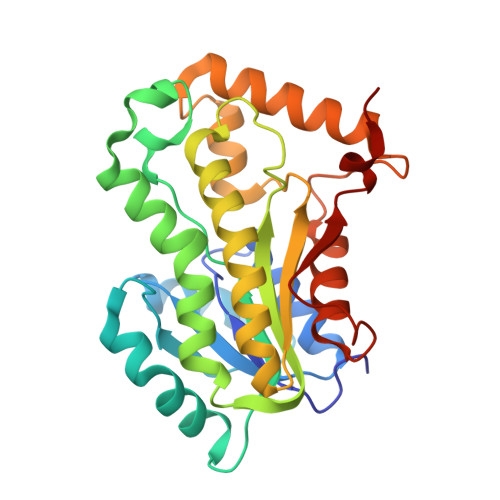
7E48

Crystal structure of InhA in complex with 3-nitropropanoic acid inhibitor


Experimental Data Snapshot

  • Method: X-RAY DIFFRACTION
  • Resolution: 2.50 Å
  • R-Value Free: 0.220 
  • R-Value Work: 0.185 

wwPDB Validation   3D Report Full Report


Ligand Structure Quality Assessment 


This is version 1.2 of the entry. See complete history


Literature

Inhibition of Mycobacterium tuberculosis InhA by 3-nitropropanoic acid.

Songsiriritthigul, C.Hanwarinroj, C.Pakamwong, B.Srimanote, P.Suttipanta, N.Sureram, S.Suttisintong, K.Kamsri, P.Punkvang, A.Spencer, J.Kittakoop, P.Pungpo, P.

(2022) Proteins 90: 898-904

  • DOI: https://doi.org/10.1002/prot.26268
  • Primary Citation of Related Structures:  
    7E48

  • PubMed Abstract: 

    3-Nitropropanoic acid (3NP), a bioactive fungal natural product, was previously demonstrated to inhibit growth of Mycobacterium tuberculosis. Here we demonstrate that 3NP inhibits the 2-trans-enoyl-acyl carrier protein reductase (InhA) from Mycobacterium tuberculosis with an IC 50 value of 71 μM, and present the crystal structure of the ternary InhA-NAD + -3NP complex. The complex contains the InhA substrate-binding loop in an ordered, open conformation with Tyr158, a catalytically important residue whose orientation defines different InhA substrate/inhibitor complex conformations, in the "out" position. 3NP occupies a hydrophobic binding site adjacent to the NAD + cofactor and close to that utilized by the diphenyl ether triclosan, but binds predominantly via electrostatic and water-mediated hydrogen-bonding interactions with the protein backbone and NAD + cofactor. The identified mode of 3NP binding provides opportunities to improve inhibitory activity toward InhA.


  • Organizational Affiliation

    Synchrotron Light Research Institute (Public Organization), Nakhon Ratchasima, Thailand.


Macromolecules
Find similar proteins by:  (by identity cutoff)  |  3D Structure
Entity ID: 1
MoleculeChains Sequence LengthOrganismDetailsImage
Enoyl-[acyl-carrier-protein] reductase [NADH]
A, B, C, D
269Mycobacterium tuberculosis H37RvMutation(s): 0 
Gene Names: inhARv1484MTCY277.05
EC: 1.3.1.9
UniProt
Find proteins for P9WGR1 (Mycobacterium tuberculosis (strain ATCC 25618 / H37Rv))
Explore P9WGR1 
Go to UniProtKB:  P9WGR1
Entity Groups  
Sequence Clusters30% Identity50% Identity70% Identity90% Identity95% Identity100% Identity
UniProt GroupP9WGR1
Sequence Annotations
Expand
  • Reference Sequence
Small Molecules
Ligands 3 Unique
IDChains Name / Formula / InChI Key2D Diagram3D Interactions
NAD
Query on NAD

Download Ideal Coordinates CCD File 
E [auth A],
G [auth B],
J [auth C],
L [auth D]
NICOTINAMIDE-ADENINE-DINUCLEOTIDE
C21 H27 N7 O14 P2
BAWFJGJZGIEFAR-NNYOXOHSSA-N
3NP
Query on 3NP

Download Ideal Coordinates CCD File 
F [auth A],
H [auth B],
K [auth C],
M [auth D]
3-NITROPROPANOIC ACID
C3 H5 N O4
WBLZUCOIBUDNBV-UHFFFAOYSA-N
GOL
Query on GOL

Download Ideal Coordinates CCD File 
I [auth B]GLYCEROL
C3 H8 O3
PEDCQBHIVMGVHV-UHFFFAOYSA-N
Experimental Data & Validation

Experimental Data

  • Method: X-RAY DIFFRACTION
  • Resolution: 2.50 Å
  • R-Value Free: 0.220 
  • R-Value Work: 0.185 
  • Space Group: P 32
Unit Cell:
Length ( Å )Angle ( ˚ )
a = 96.39α = 90
b = 96.39β = 90
c = 139.5γ = 120
Software Package:
Software NamePurpose
REFMACrefinement
MOSFLMdata reduction
SCALAdata scaling
PHASERphasing

Structure Validation

View Full Validation Report


Ligand Structure Quality Assessment 


Entry History 

Deposition Data

Revision History  (Full details and data files)

  • Version 1.0: 2021-11-24
    Type: Initial release
  • Version 1.1: 2022-02-23
    Changes: Database references
  • Version 1.2: 2023-11-29
    Changes: Data collection, Derived calculations, Refinement description