7DVV

Heme sensor protein PefR from Streptococcus agalactiae bound to operator DNA (28-mer)


Experimental Data Snapshot

  • Method: X-RAY DIFFRACTION
  • Resolution: 2.49 Å
  • R-Value Free: 0.246 
  • R-Value Work: 0.217 
  • R-Value Observed: 0.219 

wwPDB Validation   3D Report Full Report


This is version 1.1 of the entry. See complete history


Literature

Heme controls the structural rearrangement of its sensor protein mediating the hemolytic bacterial survival.

Nishinaga, M.Sugimoto, H.Nishitani, Y.Nagai, S.Nagatoishi, S.Muraki, N.Tosha, T.Tsumoto, K.Aono, S.Shiro, Y.Sawai, H.

(2021) Commun Biol 4: 467-467

  • DOI: https://doi.org/10.1038/s42003-021-01987-5
  • Primary Citation of Related Structures:  
    7DVR, 7DVS, 7DVT, 7DVU, 7DVV

  • PubMed Abstract: 

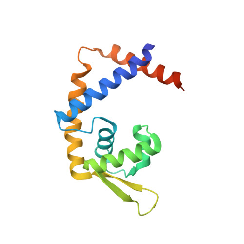
    Hemes (iron-porphyrins) are critical for biological processes in all organisms. Hemolytic bacteria survive by acquiring b-type heme from hemoglobin in red blood cells from their animal hosts. These bacteria avoid the cytotoxicity of excess heme during hemolysis by expressing heme-responsive sensor proteins that act as transcriptional factors to regulate the heme efflux system in response to the cellular heme concentration. Here, the underlying regulatory mechanisms were investigated using crystallographic, spectroscopic, and biochemical studies to understand the structural basis of the heme-responsive sensor protein PefR from Streptococcus agalactiae, a causative agent of neonatal life-threatening infections. Structural comparison of heme-free PefR, its complex with a target DNA, and heme-bound PefR revealed that unique heme coordination controls a >20 Å structural rearrangement of the DNA binding domains to dissociate PefR from the target DNA. We also found heme-bound PefR stably binds exogenous ligands, including carbon monoxide, a by-product of the heme degradation reaction.


  • Organizational Affiliation

    Graduate School of Life Science, University of Hyogo, Ako, Hyogo, Japan.


Macromolecules

Find similar proteins by:  (by identity cutoff)  |  3D Structure
Entity ID: 1
MoleculeChains Sequence LengthOrganismDetailsImage
HTH marR-type domain-containing protein
A, B
153Streptococcus agalactiae NEM316Mutation(s): 0 
Gene Names: gbs1402
UniProt
Find proteins for Q8E4J9 (Streptococcus agalactiae serotype III (strain NEM316))
Explore Q8E4J9 
Go to UniProtKB:  Q8E4J9
Entity Groups  
Sequence Clusters30% Identity50% Identity70% Identity90% Identity95% Identity100% Identity
UniProt GroupQ8E4J9
Sequence Annotations
Expand
  • Reference Sequence
Find similar nucleic acids by:  (by identity cutoff)  |  3D Structure
Entity ID: 2
MoleculeChains LengthOrganismImage
DNA (28-MER)C [auth L]28Streptococcus agalactiae NEM316
Sequence Annotations
Expand
  • Reference Sequence
Find similar nucleic acids by:  (by identity cutoff)  |  3D Structure
Entity ID: 3
MoleculeChains LengthOrganismImage
DNA (28-MER)D [auth M]28Streptococcus agalactiae NEM316
Sequence Annotations
Expand
  • Reference Sequence
Experimental Data & Validation

Experimental Data

  • Method: X-RAY DIFFRACTION
  • Resolution: 2.49 Å
  • R-Value Free: 0.246 
  • R-Value Work: 0.217 
  • R-Value Observed: 0.219 
  • Space Group: P 32 1 2
Unit Cell:
Length ( Å )Angle ( ˚ )
a = 100.973α = 90
b = 100.973β = 90
c = 128.989γ = 120
Software Package:
Software NamePurpose
REFMACrefinement
XSCALEdata scaling
PHASERphasing
PDB_EXTRACTdata extraction
Cootmodel building
XDSdata reduction

Structure Validation

View Full Validation Report


Entry History & Funding Information

Deposition Data


Funding OrganizationLocationGrant Number
Japan Society for the Promotion of Science (JSPS)JapanJP26220807
Japan Society for the Promotion of Science (JSPS)JapanJP18H02396
Japan Society for the Promotion of Science (JSPS)JapanJP18K05321

Revision History  (Full details and data files)

  • Version 1.0: 2021-09-29
    Type: Initial release
  • Version 1.1: 2023-11-29
    Changes: Data collection, Refinement description