7DVD

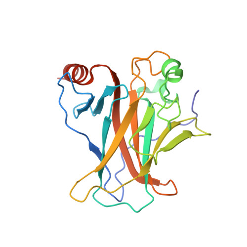
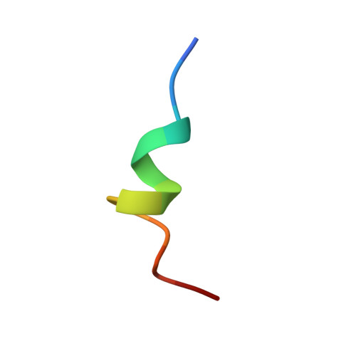
The crystal structure of p53 DNA binding domain and PUMA complex


Experimental Data Snapshot

  • Method: X-RAY DIFFRACTION
  • Resolution: 2.59 Å
  • R-Value Free: 0.318 
  • R-Value Work: 0.270 
  • R-Value Observed: 0.273 

wwPDB Validation   3D Report Full Report


This is version 1.1 of the entry. See complete history


Literature

Structural basis of the p53 DNA binding domain and PUMA complex.

Han, C.W.Lee, H.N.Jeong, M.S.Park, S.Y.Jang, S.B.

(2021) Biochem Biophys Res Commun 548: 39-46

  • DOI: https://doi.org/10.1016/j.bbrc.2021.02.049
  • Primary Citation of Related Structures:  
    7DVD

  • PubMed Abstract: 

    PUMA (p53-upregulated modulator of apoptosis) is localized in mitochondria and a direct target in p53-mediated apoptosis. p53 elicits mitochondrial apoptosis via transcription-dependent and independent mechanisms. p53 is known to induce apoptosis via the transcriptional induction of PUMA, which encodes proapoptotic BH3-only members of the Bcl-2 protein family. However, the transcription-independent mechanisms of human PUMA remain poorly defined. For example, it is not known whether PUMA interacts directly with the DNA binding domain (DBD: residues 92-293) of p53 in vitro. Here, the structure of the complex between the DBD of p53 and PUMA peptide was elucidated by X-ray crystallography. Isothermal titration calorimetry showed that PUMA peptide binds strongly with p53 DBD, and the crystal structure of p53-PUMA peptide complex revealed it contains four molecules of p53 DBD and one PUMA peptide per asymmetric unit in space group P 1 . PUMA peptide bound to the N-terminal residues of p53 DBD. A cell proliferation assay demonstrated PUMA peptide inhibited the growth of a lung cancer cell line. These results contribute to understanding of the mechanism responsible for p53-mediated apoptosis.


  • Organizational Affiliation

    Department of Molecular Biology, College of Natural Sciences, Pusan National University, 2Busandaehak-ro 63beon-gil, Geumjeong-gu, Busan, 46241, Republic of Korea.


Macromolecules
Find similar proteins by:  (by identity cutoff)  |  3D Structure
Entity ID: 1
MoleculeChains Sequence LengthOrganismDetailsImage
Cellular tumor antigen p53
A, B, C, D
205Homo sapiensMutation(s): 0 
Gene Names: TP53P53
UniProt & NIH Common Fund Data Resources
Find proteins for P04637 (Homo sapiens)
Explore P04637 
Go to UniProtKB:  P04637
PHAROS:  P04637
GTEx:  ENSG00000141510 
Entity Groups  
Sequence Clusters30% Identity50% Identity70% Identity90% Identity95% Identity100% Identity
UniProt GroupP04637
Sequence Annotations
Expand
  • Reference Sequence

Find similar proteins by:  Sequence   |   3D Structure  

Entity ID: 2
MoleculeChains Sequence LengthOrganismDetailsImage
Bcl-2-binding component 3, isoforms 1/215Homo sapiensMutation(s): 0 
UniProt & NIH Common Fund Data Resources
Find proteins for Q9BXH1 (Homo sapiens)
Explore Q9BXH1 
Go to UniProtKB:  Q9BXH1
PHAROS:  Q9BXH1
GTEx:  ENSG00000105327 
Entity Groups  
Sequence Clusters30% Identity50% Identity70% Identity90% Identity95% Identity100% Identity
UniProt GroupQ9BXH1
Sequence Annotations
Expand
  • Reference Sequence
Experimental Data & Validation

Experimental Data

  • Method: X-RAY DIFFRACTION
  • Resolution: 2.59 Å
  • R-Value Free: 0.318 
  • R-Value Work: 0.270 
  • R-Value Observed: 0.273 
  • Space Group: P 1
Unit Cell:
Length ( Å )Angle ( ˚ )
a = 68.612α = 89.939
b = 73.411β = 89.823
c = 84.408γ = 89.996
Software Package:
Software NamePurpose
HKL-2000data collection
PHENIXdata processing
Cootdata scaling
REFMACrefinement
HKL-2000data reduction

Structure Validation

View Full Validation Report


Entry History & Funding Information

Deposition Data


Funding OrganizationLocationGrant Number
National Research Foundation (NRF, Korea)Korea, Republic Of2018R1D1A1B07043701

Revision History  (Full details and data files)

  • Version 1.0: 2021-08-04
    Type: Initial release
  • Version 1.1: 2023-11-29
    Changes: Data collection, Database references, Refinement description