Nicking mechanism underlying the DNA phosphorothioate-sensing antiphage defense by SspE.
Gao, H., Gong, X., Zhou, J., Zhang, Y., Duan, J., Wei, Y., Chen, L., Deng, Z., Wang, J., Chen, S., Wu, G., Wang, L.(2022) Nat Commun 13: 6773-6773
- PubMed: 36351933 Search on PubMedSearch on PubMed Central
- DOI: https://doi.org/10.1038/s41467-022-34505-0
- Primary Citation Related Structures:
7DRI, 7DRR, 7DRS - PubMed Abstract:
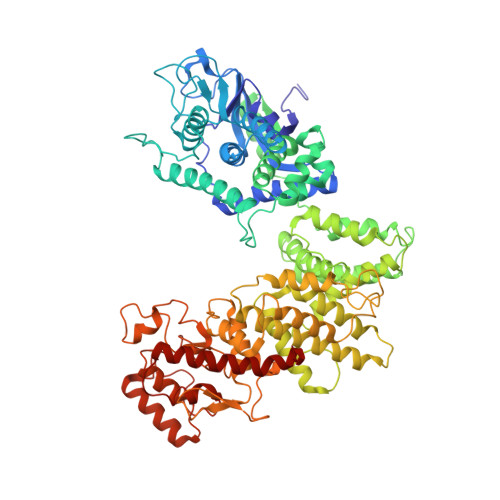
DNA phosphorothioate (PT) modification, with a nonbridging phosphate oxygen substituted by sulfur, represents a widespread epigenetic marker in prokaryotes and provides protection against genetic parasites. In the PT-based defense system Ssp, SspABCD confers a single-stranded PT modification of host DNA in the 5'-C PS CA-3' motif and SspE impedes phage propagation. SspE relies on PT modification in host DNA to exert antiphage activity. Here, structural and biochemical analyses reveal that SspE is preferentially recruited to PT sites mediated by the joint action of its N-terminal domain (NTD) hydrophobic cavity and C-terminal domain (CTD) DNA binding region. PT recognition enlarges the GTP-binding pocket, thereby increasing GTP hydrolysis activity, which subsequently triggers a conformational switch of SspE from a closed to an open state. The closed-to-open transition promotes the dissociation of SspE from self PT-DNA and turns on the DNA nicking nuclease activity of CTD, enabling SspE to accomplish self-nonself discrimination and limit phage predation, even when only a small fraction of modifiable consensus sequences is PT-protected in a bacterial genome.
- Department of Gastroenterology, TaiKang Center for Life and Medical Sciences, Zhongnan Hospital of Wuhan University, School of Pharmaceutical Sciences, Wuhan University, Wuhan, 430071, China.
Organizational Affiliation: