7DPY | pdb_00007dpy

Structure of Brucella abortus PhiA


Experimental Data Snapshot

  • Method: X-RAY DIFFRACTION
  • Resolution: 1.80 Å
  • R-Value Free: 
    0.231 (Depositor), 0.235 (DCC) 
  • R-Value Work: 
    0.199 (Depositor), 0.202 (DCC) 
  • R-Value Observed: 
    0.201 (Depositor) 

Starting Model: experimental
View more details

wwPDB Validation 3D Report Full Report

Validation slider image for 7DPY

This is version 1.4 of the entry. See complete history

Literature

Structure and Function of the Autolysin SagA in the Type IV Secretion System of Brucella abortus .

Hyun, Y.Baek, Y.Lee, C.Ki, N.Ahn, J.Ryu, S.Ha, N.C.

(2021) Mol Cells 44: 517-528

  • DOI: https://doi.org/10.14348/molcells.2021.0011
  • Primary Citation Related Structures: 
    7DNP, 7DPY

  • PubMed Abstract: 

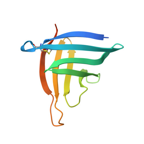
    A recent genetic study with Brucella abortus revealed the secretion activator gene A (SagA) as an autolysin component creating pores in the peptidoglycan (PGN) layer for the type IV secretion system (T4SS) and peptidoglycan hydrolase inhibitor A (PhiA) as an inhibitor of SagA. In this study, we determined the crystal structures of both SagA and PhiA. Notably, the SagA structure contained a PGN fragment in a space between the N- and C-terminal domains, showing the substrate-dependent hinge motion of the domains. The purified SagA fully hydrolyzed the meso-diaminopimelic acid (DAP)-type PGN, showing a higher activity than hen egg-white lysozyme. The PhiA protein exhibiting tetrameric assembly failed to inhibit SagA activity in our experiments. Our findings provide implications for the molecular basis of the SagA-PhiA system of B. abortus . The development of inhibitors of SagA would further contribute to controlling brucellosis by attenuating the function of T4SS, the major virulence factor of Brucella .


  • Organizational Affiliation
    • Department of Agricultural Biotechnology, Research Institute for Agriculture and Life Sciences, Center for Food and Bioconvergence, CALS, Seoul National University, Seoul 08826, Korea.

Macromolecule Content 

  • Total Structure Weight: 24.54 kDa 
  • Atom Count: 1,511 
  • Modeled Residue Count: 179 
  • Deposited Residue Count: 218 
  • Unique protein chains: 1

Macromolecules

Find similar proteins by:|  3D Structure
Entity ID: 1
MoleculeChains  Sequence LengthOrganismDetailsImage
Brucella Abortus PhiA
A, B
109Brucella abortus bv. 1 str. 9-941Mutation(s): 0 
Gene Names: BruAb1_0102
UniProt
Find proteins for Q57FR6 (Brucella abortus biovar 1 (strain 9-941))
Explore Q57FR6 
Go to UniProtKB:  Q57FR6
Entity Groups
Sequence Clusters30% Identity50% Identity70% Identity90% Identity95% Identity100% Identity
UniProt GroupQ57FR6
Sequence Annotations
Expand
Reference Sequence

Experimental Data & Validation

Experimental Data

  • Method: X-RAY DIFFRACTION
  • Resolution: 1.80 Å
  • R-Value Free:  0.231 (Depositor), 0.235 (DCC) 
  • R-Value Work:  0.199 (Depositor), 0.202 (DCC) 
  • R-Value Observed: 0.201 (Depositor) 
Space Group: P 21 21 2
Unit Cell:
Length ( Å )Angle ( ˚ )
a = 66.155α = 90
b = 51.156β = 90
c = 62.303γ = 90
Software Package:
Software NamePurpose
PHENIXrefinement
HKL-2000data reduction
HKL-2000data scaling
PHENIXphasing

Structure Validation

View Full Validation Report


Entry History 

Deposition Data

Revision History  (Full details and data files)

  • Version 1.0: 2021-06-23
    Type: Initial release
  • Version 1.1: 2021-08-11
    Changes: Database references
  • Version 1.2: 2021-10-13
    Changes: Database references
  • Version 1.3: 2023-11-29
    Changes: Data collection, Refinement description
  • Version 1.4: 2024-11-13
    Changes: Structure summary