7DDX

Crystal structure of KANK1 S1179F mutant in complex wtih eIF4A1


Experimental Data Snapshot

  • Method: X-RAY DIFFRACTION
  • Resolution: 2.50 Å
  • R-Value Free: 0.216 
  • R-Value Work: 0.191 
  • R-Value Observed: 0.192 

wwPDB Validation   3D Report Full Report


This is version 1.1 of the entry. See complete history


Literature

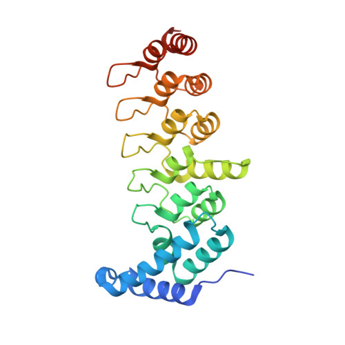
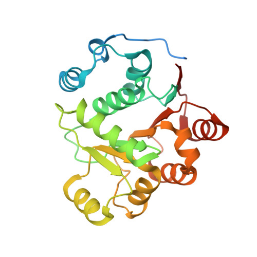
Nephrotic-syndrome-associated mutation of KANK2 induces pathologic binding competition with physiological interactor KIF21A.

Xu, Y.Guo, C.Pan, W.Zhao, C.Ding, Y.Xie, X.Wei, Z.Sun, Y.Yu, C.

(2021) J Biol Chem 297: 100958-100958

  • DOI: https://doi.org/10.1016/j.jbc.2021.100958
  • Primary Citation of Related Structures:  
    7DDX

  • PubMed Abstract: 

    Nephrotic syndrome (NS) is a common kidney disorder caused by dysfunction of the glomerular filtration barrier. Some genetic mutations identified in NS patients cause amino acid substitutions of kidney ankyrin repeat-containing (KANK) proteins, which are scaffold proteins that regulate actin polymerization, microtubule targeting, and cell adhesion via binding to various molecules, including the kinesin motor protein KIF21A. However, the mechanisms by which these mutations lead to NS are unclear. Here, we unexpectedly found that the eukaryotic translation initiation factor 4A1 (eIF4A1) interacts with an NS-associated KANK2 mutant (S684F) but not the wild-type protein. Biochemical and structural analyses revealed that the pathological mutation induces abnormal binding of eIF4A1 to KANK2 at the physiological KIF21A-binding site. Competitive binding assays further indicated that eIF4A1 can compete with KIF21A to interact with the S684F mutant of KANK2. In cultured mouse podocytes, this S684F mutant interfered with the KANK2/KIF21A interaction by binding to eIF4A1, and failed to rescue the focal adhesion or cell adhesion that had been reduced or morphologically changed by KANK2 knockout. These structural, biochemical, and cellular results not only provide mechanistic explanations for the podocyte defects caused by the S684F mutation, but also show how a gain-of-binding mutation can lead to a loss-of-function effect.


  • Organizational Affiliation

    Department of Biology, School of Life Sciences, Southern University of Science and Technology, Shenzhen, Guangdong, China.


Macromolecules
Find similar proteins by:  (by identity cutoff)  |  3D Structure
Entity ID: 1
MoleculeChains Sequence LengthOrganismDetailsImage
KN motif and ankyrin repeat domains 1257Mus musculusMutation(s): 1 
Gene Names: Kank1
UniProt & NIH Common Fund Data Resources
Find proteins for E9Q238 (Mus musculus)
Explore E9Q238 
Go to UniProtKB:  E9Q238
IMPC:  MGI:2147707
Entity Groups  
Sequence Clusters30% Identity50% Identity70% Identity90% Identity95% Identity100% Identity
UniProt GroupE9Q238
Sequence Annotations
Expand
  • Reference Sequence
Find similar proteins by:  (by identity cutoff)  |  3D Structure
Entity ID: 2
MoleculeChains Sequence LengthOrganismDetailsImage
Eukaryotic initiation factor 4A-I242Mus musculusMutation(s): 0 
Gene Names: Eif4a1Ddx2aEif4a
EC: 3.6.4.13
UniProt
Find proteins for P60843 (Mus musculus)
Explore P60843 
Go to UniProtKB:  P60843
Entity Groups  
Sequence Clusters30% Identity50% Identity70% Identity90% Identity95% Identity100% Identity
UniProt GroupP60843
Sequence Annotations
Expand
  • Reference Sequence
Experimental Data & Validation

Experimental Data

  • Method: X-RAY DIFFRACTION
  • Resolution: 2.50 Å
  • R-Value Free: 0.216 
  • R-Value Work: 0.191 
  • R-Value Observed: 0.192 
  • Space Group: P 31 2 1
Unit Cell:
Length ( Å )Angle ( ˚ )
a = 137.628α = 90
b = 137.628β = 90
c = 57.54γ = 120
Software Package:
Software NamePurpose
PHENIXrefinement
PDB_EXTRACTdata extraction
HKL-2000data reduction
HKL-2000data scaling
PHASERphasing

Structure Validation

View Full Validation Report


Entry History & Funding Information

Deposition Data


Funding OrganizationLocationGrant Number
National Natural Science Foundation of China (NSFC)China31770791
National Natural Science Foundation of China (NSFC)China31971131

Revision History  (Full details and data files)

  • Version 1.0: 2021-09-08
    Type: Initial release
  • Version 1.1: 2023-11-29
    Changes: Data collection, Refinement description