7DCE

Cryo-EM structure of human XKR8-basigin complex bound to Fab fragment


Experimental Data Snapshot

  • Method: ELECTRON MICROSCOPY
  • Resolution: 3.80 Å
  • Aggregation State: PARTICLE 
  • Reconstruction Method: SINGLE PARTICLE 

wwPDB Validation   3D Report Full Report


This is version 2.0 of the entry. See complete history


Literature

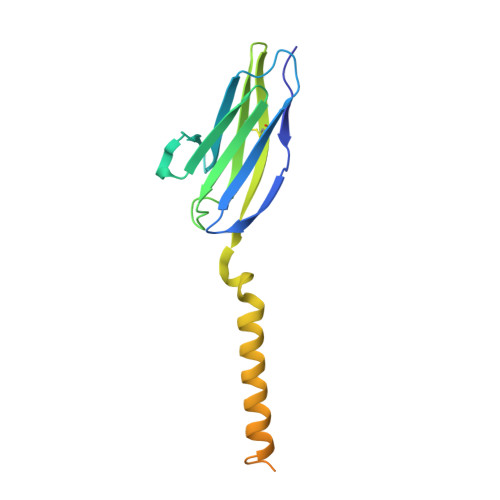
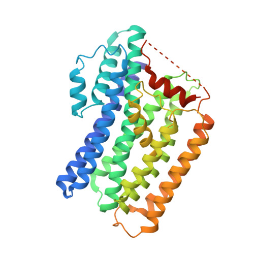
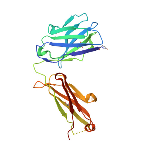
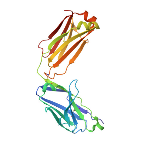
The tertiary structure of the human Xkr8-Basigin complex that scrambles phospholipids at plasma membranes.

Sakuragi, T.Kanai, R.Tsutsumi, A.Narita, H.Onishi, E.Nishino, K.Miyazaki, T.Baba, T.Kosako, H.Nakagawa, A.Kikkawa, M.Toyoshima, C.Nagata, S.

(2021) Nat Struct Mol Biol 28: 825-834

  • DOI: https://doi.org/10.1038/s41594-021-00665-8
  • Primary Citation of Related Structures:  
    7D9Z, 7DAA, 7DCE

  • PubMed Abstract: 

    Xkr8-Basigin is a plasma membrane phospholipid scramblase activated by kinases or caspases. We combined cryo-EM and X-ray crystallography to investigate its structure at an overall resolution of 3.8 Å. Its membrane-spanning region carrying 22 charged amino acids adopts a cuboid-like structure stabilized by salt bridges between hydrophilic residues in transmembrane helices. Phosphatidylcholine binding was observed in a hydrophobic cleft on the surface exposed to the outer leaflet of the plasma membrane. Six charged residues placed from top to bottom inside the molecule were essential for scrambling phospholipids in inward and outward directions, apparently providing a pathway for their translocation. A tryptophan residue was present between the head group of phosphatidylcholine and the extracellular end of the path. Its mutation to alanine made the Xkr8-Basigin complex constitutively active, indicating that it plays a vital role in regulating its scramblase activity. The structure of Xkr8-Basigin provides insights into the molecular mechanisms underlying phospholipid scrambling.


  • Organizational Affiliation

    Laboratory of Biochemistry and Immunology, World Premier International Immunology Frontier Research Center, Osaka University, Suita, Japan.


Macromolecules
Find similar proteins by:  (by identity cutoff)  |  3D Structure
Entity ID: 1
MoleculeChains Sequence LengthOrganismDetailsImage
Isoform 2 of BasiginA [auth B]176Homo sapiensMutation(s): 2 
Gene Names: BSGUNQ6505/PRO21383
Membrane Entity: Yes 
UniProt & NIH Common Fund Data Resources
Find proteins for P35613 (Homo sapiens)
Explore P35613 
Go to UniProtKB:  P35613
PHAROS:  P35613
GTEx:  ENSG00000172270 
Entity Groups  
Sequence Clusters30% Identity50% Identity70% Identity90% Identity95% Identity100% Identity
UniProt GroupP35613
Sequence Annotations
Expand
  • Reference Sequence
Find similar proteins by:  (by identity cutoff)  |  3D Structure
Entity ID: 2
MoleculeChains Sequence LengthOrganismDetailsImage
XK-related protein 8B [auth X]405Homo sapiensMutation(s): 0 
Gene Names: XKR8XRG8
Membrane Entity: Yes 
UniProt & NIH Common Fund Data Resources
Find proteins for Q9H6D3 (Homo sapiens)
Explore Q9H6D3 
Go to UniProtKB:  Q9H6D3
GTEx:  ENSG00000158156 
Entity Groups  
Sequence Clusters30% Identity50% Identity70% Identity90% Identity95% Identity100% Identity
UniProt GroupQ9H6D3
Sequence Annotations
Expand
  • Reference Sequence
Find similar proteins by:  (by identity cutoff)  |  3D Structure
Entity ID: 3
MoleculeChains Sequence LengthOrganismDetailsImage
Heavy chain of Fab fragmentC [auth H]217Oryctolagus cuniculusMutation(s): 0 
Entity Groups  
Sequence Clusters30% Identity50% Identity70% Identity90% Identity95% Identity100% Identity
Sequence Annotations
Expand
  • Reference Sequence
Find similar proteins by:  (by identity cutoff)  |  3D Structure
Entity ID: 4
MoleculeChains Sequence LengthOrganismDetailsImage
Light chain of Fab fragmentD [auth L]218Oryctolagus cuniculusMutation(s): 0 
Entity Groups  
Sequence Clusters30% Identity50% Identity70% Identity90% Identity95% Identity100% Identity
Sequence Annotations
Expand
  • Reference Sequence
Small Molecules
Ligands 1 Unique
IDChains Name / Formula / InChI Key2D Diagram3D Interactions
DLP (Subject of Investigation/LOI)
Query on DLP

Download Ideal Coordinates CCD File 
E [auth X]1,2-DILINOLEOYL-SN-GLYCERO-3-PHOSPHOCHOLINE
C44 H80 N O8 P
FVXDQWZBHIXIEJ-LNDKUQBDSA-N
Modified Residues  1 Unique
IDChains TypeFormula2D DiagramParent
PCA
Query on PCA
C [auth H]L-PEPTIDE LINKINGC5 H7 N O3GLN
Experimental Data & Validation

Experimental Data

  • Method: ELECTRON MICROSCOPY
  • Resolution: 3.80 Å
  • Aggregation State: PARTICLE 
  • Reconstruction Method: SINGLE PARTICLE 

Structure Validation

View Full Validation Report


Entry History & Funding Information

Deposition Data


Funding OrganizationLocationGrant Number
Japan Society for the Promotion of Science (JSPS)Japan15H05785
Japan Society for the Promotion of Science (JSPS)Japan20K15731
Japan Science and TechnologyJapanJPMJCR14M1
Japan Science and TechnologyJapanJPMJCR14M4
Japan Agency for Medical Research and Development (AMED)JapanJP17am010172
Japan Agency for Medical Research and Development (AMED)JapanJP18am0101072
Japan Agency for Medical Research and Development (AMED)JapanJP19am0101072

Revision History  (Full details and data files)

  • Version 1.0: 2021-10-20
    Type: Initial release
  • Version 2.0: 2024-01-24
    Type: Coordinate replacement
    Reason: Polymer geometry
    Changes: Advisory, Atomic model, Data collection, Derived calculations, Source and taxonomy, Structure summary