


Structure of 6-hydroxy-3-succinoyl-pyridine 3-monooxygenase (HspB) from Pseudomonas putida S16
Liu, G.Q., Liu, G.Q.To be published.
Experimental Data Snapshot
Starting Model: experimental
View more details
wwPDB Validation 3D Report Full Report

Entity ID: 1 | |||||
|---|---|---|---|---|---|
| Molecule | Chains | Sequence Length | Organism | Details | Image |
| 6-hydroxy-3-succinoylpyridine 3-monooxygenase HspB | 393 | Pseudomonas putida S16 | Mutation(s): 0 Gene Names: hspB, PPS_4061 EC: 1.14.13.163 | 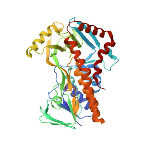 | |
UniProt | |||||
Entity Groups | |||||
| Sequence Clusters | 30% Identity50% Identity70% Identity90% Identity95% Identity100% Identity | ||||
| UniProt Group | F8G0M4 | ||||
Sequence AnnotationsExpand | |||||
Reference Sequence | |||||
| Length ( Å ) | Angle ( ˚ ) |
|---|---|
| a = 67.417 | α = 90 |
| b = 83.779 | β = 114.95 |
| c = 79.56 | γ = 90 |
| Software Name | Purpose |
|---|---|
| HKL-2000 | data scaling |
| PHENIX | refinement |
| PDB_EXTRACT | data extraction |
| HKL-3000 | data reduction |
| HKL-3000 | phasing |
| Funding Organization | Location | Grant Number |
|---|---|---|
| National Science Foundation (NSF, China) | China | -- |


