Hypomorphic mutations in human DNA ligase IV lead to compromised DNA binding efficiency, hydrophobicity and thermal stability.
Maddi, E.R., Raghavan, S.C., Natesh, R.(2021) Protein Eng Des Sel 34
- PubMed: 33586762 Search on PubMed
- DOI: https://doi.org/10.1093/protein/gzab001
- Primary Citation Related Structures:
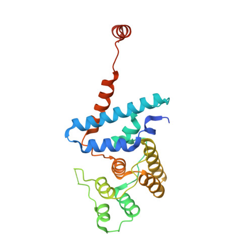
7D9K, 7D9Y - PubMed Abstract:
Studies have shown that Lig4 syndrome mutations in DNA ligase IV (LigIV) are compromised in its function with residual level of double strand break ligation activity in vivo. It was speculated that Lig4 syndrome mutations adversely affect protein folding and stability. Though there are crystal structures of LigIV, there are no reports of crystal structures of Lig4 syndrome mutants and their biophysical characterization to date. Here, we have examined the conformational states, thermal stability, hydrophobicity and DNA binding efficiency of human DNA LigIV wild type and its hypomorphic mutants by far-UV circular dichroism, tyrosine and tryptophan fluorescence, and 1-anilino-8-naphthalene-sulfonate binding, dynamic light scattering, size exclusion chromatography, multi-angle light scattering and electrophoretic mobility shift assay. We show here that LigIV hypomorphic mutants have reduced DNA-binding efficiency, a shift in secondary structure content from the helical to random coil, marginal reduction in their thermal stability and increased hydrophobicity as compared to the wild-type LigIV.
- School of Biology, Indian Institute of Science Education and Research Thiruvananthapuram, Thiruvananthapuram, Kerala 695551, India.
Organizational Affiliation: