7D0C

S protein of SARS-CoV-2 in complex bound with P5A-3A1


Experimental Data Snapshot

  • Method: ELECTRON MICROSCOPY
  • Resolution: 3.40 Å
  • Aggregation State: PARTICLE 
  • Reconstruction Method: SINGLE PARTICLE 

wwPDB Validation   3D Report Full Report


This is version 1.2 of the entry. See complete history


Literature

Structural basis for bivalent binding and inhibition of SARS-CoV-2 infection by human potent neutralizing antibodies.

Yan, R.Wang, R.Ju, B.Yu, J.Zhang, Y.Liu, N.Wang, J.Zhang, Q.Chen, P.Zhou, B.Li, Y.Shen, Y.Zhang, S.Tian, L.Guo, Y.Xia, L.Zhong, X.Cheng, L.Ge, X.Zhao, J.Wang, H.W.Wang, X.Zhang, Z.Zhang, L.Zhou, Q.

(2021) Cell Res 31: 517-525

  • DOI: https://doi.org/10.1038/s41422-021-00487-9
  • Primary Citation of Related Structures:  
    7CZP, 7CZQ, 7CZR, 7CZS, 7CZT, 7CZU, 7CZV, 7CZW, 7CZX, 7CZY, 7CZZ, 7D00, 7D03, 7D0B, 7D0C, 7D0D

  • PubMed Abstract: 

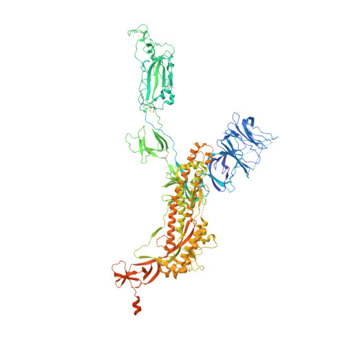
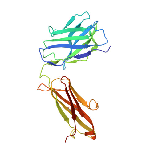
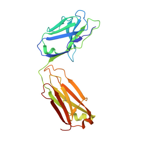
    Neutralizing monoclonal antibodies (nAbs) to severe acute respiratory syndrome coronavirus 2 (SARS-CoV-2) represent promising candidates for clinical intervention against coronavirus disease 2019 (COVID-19). We isolated a large number of nAbs from SARS-CoV-2-infected individuals capable of disrupting proper interaction between the receptor binding domain (RBD) of the viral spike (S) protein and the receptor angiotensin converting enzyme 2 (ACE2). However, the structural basis for their potent neutralizing activity remains unclear. Here, we report cryo-EM structures of the ten most potent nAbs in their native full-length IgG-form or in both IgG-form and Fab-form bound to the trimeric S protein of SARS-CoV-2. The bivalent binding of the full-length IgG is found to associate with more RBDs in the "up" conformation than the monovalent binding of Fab, perhaps contributing to the enhanced neutralizing activity of IgG and triggering more shedding of the S1 subunit from the S protein. Comparison of a large number of nAbs identified common and unique structural features associated with their potent neutralizing activities. This work provides a structural basis for further understanding the mechanism of nAbs, especially through revealing the bivalent binding and its correlation with more potent neutralization and the shedding of S1 subunit.


  • Organizational Affiliation

    Center for Infectious Disease Research, Westlake Laboratory of Life Sciences and Biomedicine, Key Laboratory of Structural Biology of Zhejiang Province, School of Life Sciences, Westlake University, 18 Shilongshan Road, Hangzhou, Zhejiang, 310024, China.


Macromolecules
Find similar proteins by:  (by identity cutoff)  |  3D Structure
Entity ID: 1
MoleculeChains Sequence LengthOrganismDetailsImage
Spike glycoprotein
A, B, C
1,283Severe acute respiratory syndrome coronavirus 2Mutation(s): 2 
Gene Names: S2
UniProt
Find proteins for P0DTC2 (Severe acute respiratory syndrome coronavirus 2)
Explore P0DTC2 
Go to UniProtKB:  P0DTC2
Entity Groups  
Sequence Clusters30% Identity50% Identity70% Identity90% Identity95% Identity100% Identity
UniProt GroupP0DTC2
Sequence Annotations
Expand
  • Reference Sequence
Find similar proteins by:  (by identity cutoff)  |  3D Structure
Entity ID: 2
MoleculeChains Sequence LengthOrganismDetailsImage
Heavy chain of P5A-3A1D [auth H],
F [auth G]
219Homo sapiensMutation(s): 0 
Entity Groups  
Sequence Clusters30% Identity50% Identity70% Identity90% Identity95% Identity100% Identity
Sequence Annotations
Expand
  • Reference Sequence
Find similar proteins by:  (by identity cutoff)  |  3D Structure
Entity ID: 3
MoleculeChains Sequence LengthOrganismDetailsImage
Light chain of P5A-3A1E [auth L],
G [auth F]
215Homo sapiensMutation(s): 0 
Entity Groups  
Sequence Clusters30% Identity50% Identity70% Identity90% Identity95% Identity100% Identity
Sequence Annotations
Expand
  • Reference Sequence
Small Molecules
Ligands 1 Unique
IDChains Name / Formula / InChI Key2D Diagram3D Interactions
NAG (Subject of Investigation/LOI)
Query on NAG

Download Ideal Coordinates CCD File 
AB [auth C]
BB [auth C]
CB [auth C]
DA [auth A]
EA [auth A]
AB [auth C],
BB [auth C],
CB [auth C],
DA [auth A],
EA [auth A],
FA [auth A],
GA [auth A],
HA [auth A],
IA [auth A],
JA [auth A],
KA [auth A],
LA [auth A],
MA [auth B],
NA [auth B],
OA [auth B],
PA [auth B],
QA [auth B],
RA [auth B],
SA [auth B],
TA [auth B],
UA [auth B],
VA [auth C],
WA [auth C],
XA [auth C],
YA [auth C],
ZA [auth C]
2-acetamido-2-deoxy-beta-D-glucopyranose
C8 H15 N O6
OVRNDRQMDRJTHS-FMDGEEDCSA-N
Experimental Data & Validation

Experimental Data

  • Method: ELECTRON MICROSCOPY
  • Resolution: 3.40 Å
  • Aggregation State: PARTICLE 
  • Reconstruction Method: SINGLE PARTICLE 
EM Software:
TaskSoftware PackageVersion
MODEL REFINEMENTPHENIX

Structure Validation

View Full Validation Report


Entry History 

Deposition Data

Revision History  (Full details and data files)

  • Version 1.0: 2021-03-10
    Type: Initial release
  • Version 1.1: 2021-04-07
    Changes: Database references
  • Version 1.2: 2021-05-19
    Changes: Database references