7CT4 | pdb_00007ct4

Crystal structure of D-amino acid oxidase from Rasamsonia emersonii strain YA


Experimental Data Snapshot

  • Method: X-RAY DIFFRACTION
  • Resolution: 2.00 Å
  • R-Value Free: 
    0.246 (Depositor), 0.233 (DCC) 
  • R-Value Work: 
    0.206 (Depositor) 
  • R-Value Observed: 
    0.208 (Depositor) 

Starting Model: experimental
View more details

wwPDB Validation 3D Report Full Report

Validation slider image for 7CT4

Ligand Structure Quality Assessment 


This is version 1.2 of the entry. See complete history

Literature

X-ray structure analysis of a unique D-amino-acid oxidase from the thermophilic fungus Rasamsonia emersonii strain YA.

Shimekake, Y.Hirato, Y.Funabashi, R.Okazaki, S.Goto, M.Furuichi, T.Suzuki, H.Kera, Y.Takahashi, S.

(2020) Acta Crystallogr F Struct Biol Commun 76: 517-523

  • DOI: https://doi.org/10.1107/S2053230X20013333
  • Primary Citation Related Structures: 
    7CT4

  • PubMed Abstract: 

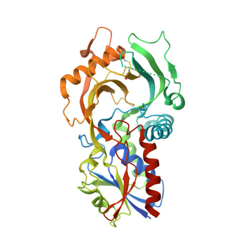
    D-Amino-acid oxidases (DAAOs) catalyze the oxidative deamination of neutral and basic D-amino acids. The DAAO from the thermophilic fungus Rasamsonia emersonii strain YA (ReDAAO) has a high thermal stability and a unique broad substrate specificity that includes the acidic D-amino acid D-Glu as well as various neutral and basic D-amino acids. In this study, ReDAAO was crystallized by the hanging-drop vapor-diffusion method and its crystal structure was determined at a resolution of 2.00 Å. The crystal structure of the enzyme revealed that unlike other DAAOs, ReDAAO forms a homotetramer and contains an intramolecular disulfide bond (Cys230-Cys285), suggesting that this disulfide bond is involved in the higher thermal stability of ReDAAO. Moreover, the structure of the active site and its vicinity in ReDAAO indicates that Arg97, Lys99, Lys114 and Ser231 are candidates for recognizing the side chain of D-Glu.


  • Organizational Affiliation
    • Department of Bioengineering, Nagaoka University of Technology, 1603-1 Kamitomioka, Nagaoka, Niigata 940-2188, Japan.

Macromolecule Content 

  • Total Structure Weight: 173.2 kDa 
  • Atom Count: 10,965 
  • Modeled Residue Count: 1,376 
  • Deposited Residue Count: 1,552 
  • Unique protein chains: 1

Macromolecules

Find similar proteins by:|  3D Structure
Entity ID: 1
MoleculeChains  Sequence LengthOrganismDetailsImage
D-amino acid oxidase
A, B, C, D
388Rasamsonia emersoniiMutation(s): 0 
Gene Names: ReDAO
EC: 1.4.3.3
UniProt
Find proteins for A0A499UB99 (Rasamsonia emersonii)
Explore A0A499UB99 
Go to UniProtKB:  A0A499UB99
Entity Groups
Sequence Clusters30% Identity50% Identity70% Identity90% Identity95% Identity100% Identity
UniProt GroupA0A499UB99
Sequence Annotations
Expand
Reference Sequence

Experimental Data & Validation

Experimental Data

  • Method: X-RAY DIFFRACTION
  • Resolution: 2.00 Å
  • R-Value Free:  0.246 (Depositor), 0.233 (DCC) 
  • R-Value Work:  0.206 (Depositor) 
  • R-Value Observed: 0.208 (Depositor) 
Space Group: P 61
Unit Cell:
Length ( Å )Angle ( ˚ )
a = 79.13α = 90
b = 79.13β = 90
c = 366.673γ = 120
Software Package:
Software NamePurpose
REFMACrefinement
XDSdata reduction
SCALAdata scaling
PDB_EXTRACTdata extraction
MOLREPphasing

Structure Validation

View Full Validation Report


Ligand Structure Quality Assessment 


Entry History 

Deposition Data

Revision History  (Full details and data files)

  • Version 1.0: 2020-11-11
    Type: Initial release
  • Version 1.1: 2023-11-29
    Changes: Data collection, Database references, Refinement description
  • Version 1.2: 2024-10-23
    Changes: Structure summary