7CHR

AcrIF9


Experimental Data Snapshot

  • Method: X-RAY DIFFRACTION
  • Resolution: 1.21 Å
  • R-Value Free: 0.195 
  • R-Value Work: 0.188 
  • R-Value Observed: 0.189 

wwPDB Validation   3D Report Full Report


This is version 1.1 of the entry. See complete history


Literature

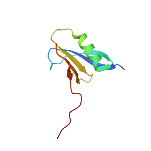
A high-resolution (1.2 angstrom ) crystal structure of the anti-CRISPR protein AcrIF9.

Kim, G.E.Lee, S.Y.Park, H.H.

(2020) FEBS Open Bio 10: 2532-2540

  • DOI: https://doi.org/10.1002/2211-5463.12986
  • Primary Citation of Related Structures:  
    7CHR

  • PubMed Abstract: 

    Prokaryotic adaptive immunity by CRISPR-Cas systems, which confer resistance to foreign genetic elements, has been used by bacteria to combat viruses. To cope, viruses evolved multiple anti-CRISPR proteins, which can inhibit system function through various mechanisms. Although the structures and mechanisms of several anti-CRISPR proteins have been elucidated, those of the AcrIF9 family have not yet been identified. To understand the molecular basis underlying AcrIF9 anti-CRISPR function, we determined the 1.2 Å crystal structure of AcrIF9. Structural and biochemical studies showed that AcrIF9 exists in monomeric form in solution and can directly interact with DNA using a positively charged cleft. Based on analysis of the structure, we suggest part of the anti-CRISPR molecular mechanism by AcrIF9.


  • Organizational Affiliation

    Department of Global Innovative Drugs, Graduate School of Chung-Ang University, Seoul, Korea.


Macromolecules
Find similar proteins by:  (by identity cutoff)  |  3D Structure
Entity ID: 1
MoleculeChains Sequence LengthOrganismDetailsImage
anti-CRISPR AcrIF976Photobacterium damselaeMutation(s): 0 
Gene Names: BST98_20680
UniProt
Find proteins for A0A2S2CJ39 (Photobacterium damselae)
Explore A0A2S2CJ39 
Go to UniProtKB:  A0A2S2CJ39
Entity Groups  
Sequence Clusters30% Identity50% Identity70% Identity90% Identity95% Identity100% Identity
UniProt GroupA0A2S2CJ39
Sequence Annotations
Expand
  • Reference Sequence
Experimental Data & Validation

Experimental Data

  • Method: X-RAY DIFFRACTION
  • Resolution: 1.21 Å
  • R-Value Free: 0.195 
  • R-Value Work: 0.188 
  • R-Value Observed: 0.189 
  • Space Group: P 21 21 21
Unit Cell:
Length ( Å )Angle ( ˚ )
a = 27.23α = 90
b = 31.35β = 90
c = 80.7γ = 90
Software Package:
Software NamePurpose
PHENIXrefinement
MxDCdata collection
XDSdata scaling
PHASERphasing
Cootmodel building

Structure Validation

View Full Validation Report


Entry History & Funding Information

Deposition Data


Funding OrganizationLocationGrant Number
National Research Foundation (NRF, Korea)Korea, Republic Of--

Revision History  (Full details and data files)

  • Version 1.0: 2020-12-23
    Type: Initial release
  • Version 1.1: 2024-03-27
    Changes: Data collection, Database references, Derived calculations