


Structure of glucose dehydrogenase at 1.33 Angstroms
Jia, S., Xu, D., Wang, W., Ran, T.To be published.
Experimental Data Snapshot
Starting Model: experimental
View more details
Entity ID: 1 | |||||
|---|---|---|---|---|---|
| Molecule | Chains | Sequence Length | Organism | Details | Image |
| glucose dehydrogenase | 355 | Serratia sp. FS14 | Mutation(s): 0 | 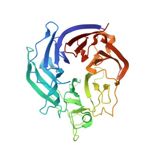 | |
UniProt | |||||
Find proteins for A0A8I3AZU9 (Serratia sp. (strain FS14)) Explore A0A8I3AZU9 Go to UniProtKB: A0A8I3AZU9 | |||||
Entity Groups | |||||
| Sequence Clusters | 30% Identity50% Identity70% Identity90% Identity95% Identity100% Identity | ||||
| UniProt Group | A0A8I3AZU9 | ||||
Sequence AnnotationsExpand | |||||
Reference Sequence | |||||
| Ligands 2 Unique | |||||
|---|---|---|---|---|---|
| ID | Chains | Name / Formula / InChI Key | 2D Diagram | 3D Interactions | |
| GOL (Subject of Investigation/LOI) Download:Ideal Coordinates CCD File | E [auth B] | GLYCEROL C3 H8 O3 PEDCQBHIVMGVHV-UHFFFAOYSA-N |  | ||
| CA (Subject of Investigation/LOI) Download:Ideal Coordinates CCD File | C [auth A], D [auth B] | CALCIUM ION Ca BHPQYMZQTOCNFJ-UHFFFAOYSA-N |  | ||
| Length ( Å ) | Angle ( ˚ ) |
|---|---|
| a = 160.622 | α = 90 |
| b = 44.64 | β = 100.096 |
| c = 99.885 | γ = 90 |
| Software Name | Purpose |
|---|---|
| REFMAC | refinement |
| HKL-3000 | data reduction |
| HKL-3000 | data scaling |
| PHASER | phasing |
| Funding Organization | Location | Grant Number |
|---|---|---|
| National Science Foundation (NSF, China) | China | 31770074 |
| National Science Foundation (NSF, China) | China | 31770050 |


