7CCC

The structure of the actin filament uncapping complex mediated by twinfilin


Experimental Data Snapshot

  • Method: X-RAY DIFFRACTION
  • Resolution: 3.20 Å
  • R-Value Free: 0.238 
  • R-Value Work: 0.186 
  • R-Value Observed: 0.192 

wwPDB Validation   3D Report Full Report


Ligand Structure Quality Assessment 


This is version 1.2 of the entry. See complete history


Literature

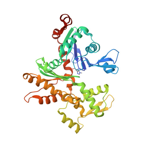
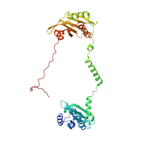
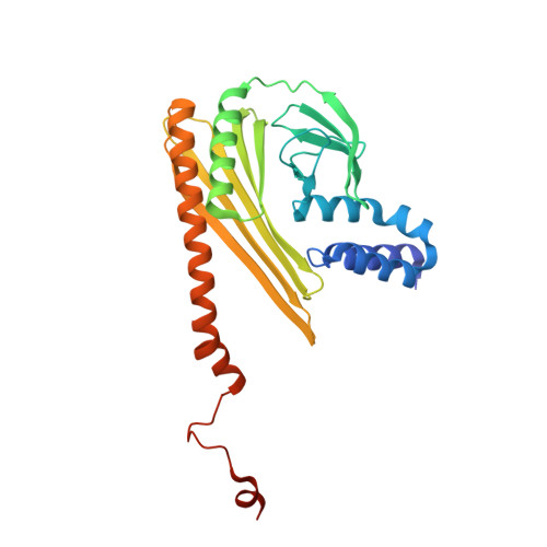
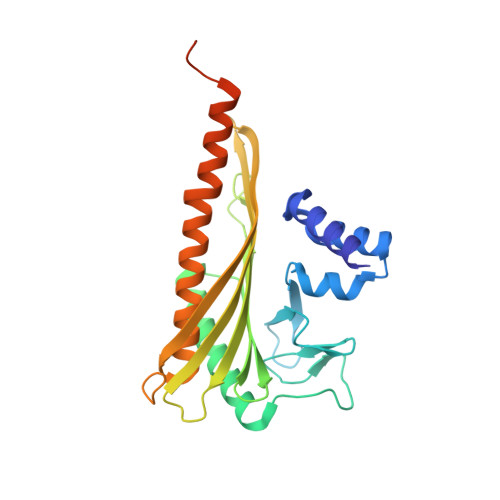
The structure of the actin filament uncapping complex mediated by twinfilin.

Mwangangi, D.M.Manser, E.Robinson, R.C.

(2021) Sci Adv 7

  • DOI: https://doi.org/10.1126/sciadv.abd5271
  • Primary Citation of Related Structures:  
    7CCC

  • PubMed Abstract: 

    Uncapping of actin filaments is essential for driving polymerization and depolymerization dynamics from capping protein-associated filaments; however, the mechanisms of uncapping leading to rapid disassembly are unknown. Here, we elucidated the x-ray crystal structure of the actin/twinfilin/capping protein complex to address the mechanisms of twinfilin uncapping of actin filaments. The twinfilin/capping protein complex binds to two G-actin subunits in an orientation that resembles the actin filament barbed end. This suggests an unanticipated mechanism by which twinfilin disrupts the stable capping of actin filaments by inducing a G-actin conformation in the two terminal actin subunits. Furthermore, twinfilin disorders critical actin-capping protein interactions, which will assist in the dissociation of capping protein, and may promote filament uncapping through a second mechanism involving V-1 competition for an actin-binding surface on capping protein. The extensive interactions with capping protein indicate that the evolutionary conserved role of twinfilin is to uncap actin filaments.


  • Organizational Affiliation

    Institute of Molecular and Cell Biology, A*STAR (Agency for Science, Technology and Research), Biopolis, Singapore 138673, Singapore.


Macromolecules
Find similar proteins by:  (by identity cutoff)  |  3D Structure
Entity ID: 1
MoleculeChains Sequence LengthOrganismDetailsImage
Actin, alpha skeletal muscle
A, E
377Oryctolagus cuniculusMutation(s): 0 
UniProt
Find proteins for P68135 (Oryctolagus cuniculus)
Explore P68135 
Go to UniProtKB:  P68135
Entity Groups  
Sequence Clusters30% Identity50% Identity70% Identity90% Identity95% Identity100% Identity
UniProt GroupP68135
Sequence Annotations
Expand
  • Reference Sequence
Find similar proteins by:  (by identity cutoff)  |  3D Structure
Entity ID: 2
MoleculeChains Sequence LengthOrganismDetailsImage
Twinfilin-1350Homo sapiensMutation(s): 0 
Gene Names: TWF1PTK9
UniProt & NIH Common Fund Data Resources
Find proteins for Q12792 (Homo sapiens)
Explore Q12792 
Go to UniProtKB:  Q12792
PHAROS:  Q12792
GTEx:  ENSG00000151239 
Entity Groups  
Sequence Clusters30% Identity50% Identity70% Identity90% Identity95% Identity100% Identity
UniProt GroupQ12792
Sequence Annotations
Expand
  • Reference Sequence
Find similar proteins by:  (by identity cutoff)  |  3D Structure
Entity ID: 3
MoleculeChains Sequence LengthOrganismDetailsImage
F-actin-capping protein subunit alpha286Mus musculusMutation(s): 1 
Gene Names: Capza1
UniProt & NIH Common Fund Data Resources
Find proteins for P47753 (Mus musculus)
Explore P47753 
Go to UniProtKB:  P47753
IMPC:  MGI:106227
Entity Groups  
Sequence Clusters30% Identity50% Identity70% Identity90% Identity95% Identity100% Identity
UniProt GroupP47753
Sequence Annotations
Expand
  • Reference Sequence
Find similar proteins by:  (by identity cutoff)  |  3D Structure
Entity ID: 4
MoleculeChains Sequence LengthOrganismDetailsImage
F-actin-capping protein subunit beta272Mus musculusMutation(s): 0 
Gene Names: Capzb
UniProt & NIH Common Fund Data Resources
Find proteins for Q923G3 (Mus musculus)
Explore Q923G3 
Go to UniProtKB:  Q923G3
IMPC:  MGI:104652
Entity Groups  
Sequence Clusters30% Identity50% Identity70% Identity90% Identity95% Identity100% Identity
UniProt GroupQ923G3
Sequence Annotations
Expand
  • Reference Sequence
Experimental Data & Validation

Experimental Data

  • Method: X-RAY DIFFRACTION
  • Resolution: 3.20 Å
  • R-Value Free: 0.238 
  • R-Value Work: 0.186 
  • R-Value Observed: 0.192 
  • Space Group: P 21 21 21
Unit Cell:
Length ( Å )Angle ( ˚ )
a = 74.759α = 90
b = 168.662β = 90
c = 176.989γ = 90
Software Package:
Software NamePurpose
PHENIXrefinement
HKL-2000data reduction
HKL-2000data scaling
PHASERphasing
PDB_EXTRACTdata extraction

Structure Validation

View Full Validation Report


Ligand Structure Quality Assessment 


Entry History & Funding Information

Deposition Data


Funding OrganizationLocationGrant Number
Human Frontier Science Program (HFSP)RGP0028/2018

Revision History  (Full details and data files)

  • Version 1.0: 2021-02-03
    Type: Initial release
  • Version 1.1: 2021-02-24
    Changes: Database references
  • Version 1.2: 2023-11-29
    Changes: Data collection, Database references, Refinement description