7C90

Crystal structure of Cytochrome CL from the marine methylotrophic bacterium Methylophaga aminisulfidivorans MPT (Ma-CytcL)


Experimental Data Snapshot

  • Method: X-RAY DIFFRACTION
  • Resolution: 2.13 Å
  • R-Value Free: 0.200 
  • R-Value Work: 0.148 
  • R-Value Observed: 0.151 

wwPDB Validation   3D Report Full Report


Ligand Structure Quality Assessment 


This is version 1.2 of the entry. See complete history


Literature

Crystal Structure of CytochromecLfrom the Aquatic Methylotrophic BacteriumMethylophaga aminisulfidivoransMPT.

Ghosh, S.Dhanasingh, I.Ryu, J.Kim, S.W.Lee, S.H.

(2020) J Microbiol Biotechnol 30: 1261-1271

  • DOI: https://doi.org/10.4014/jmb.2006.06029
  • Primary Citation of Related Structures:  
    7C90

  • PubMed Abstract: 

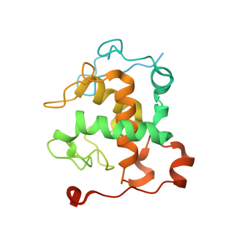
    Cytochrome c L (Cyt c L ) is an essential protein in the process of methanol oxidation in methylotrophs. It receives an electron from the pyrroloquinoline quinone (PQQ) cofactor of methanol dehydrogenase (MDH) to produce formaldehyde. The direct electron transfer mechanism between Cyt c L and MDH remains unknown due to the lack of structural information. To help gain a better understanding of the mechanism, we determined the first crystal structure of heme c containing Cyt c L from the aquatic methylotrophic bacterium Methylophaga aminisulfidivorans MP T at 2.13 Å resolution. The crystal structure of Ma -Cyt c L revealed its unique features compared to those of the terrestrial homologues. Apart from Fe in heme, three additional metal ion binding sites for Na + , Ca + , and Fe 2+ were found, wherein the ions mostly formed coordination bonds with the amino acid residues on the loop (G93-Y111) that interacts with heme. Therefore, these ions seemed to enhance the stability of heme insertion by increasing the loop's steadiness. The basic N-terminal end, together with helix α4 and loop (G126 to Y136), contributed positive charge to the region. In contrast, the acidic C-terminal end provided a negatively charged surface, yielding several electrostatic contact points with partner proteins for electron transfer. These exceptional features of Ma -Cyt c L , along with the structural information of MDH, led us to hypothesize the need for an adapter protein bridging MDH to Cyt c L within appropriate proximity for electron transfer. With this knowledge in mind, the methanol oxidation complex reconstitution in vitro could be utilized to produce metabolic intermediates at the industry level.


  • Organizational Affiliation

    Department of Cellular and Molecular Medicine, Chosun University School of Medicine, Gwangju 61452, Republic of Korea.


Macromolecules
Find similar proteins by:  (by identity cutoff)  |  3D Structure
Entity ID: 1
MoleculeChains Sequence LengthOrganismDetailsImage
Cytochrome c, mono-and diheme variant
A, B, C, D
194Methylophaga aminisulfidivorans MPMutation(s): 0 
Gene Names: MAMP_01204
UniProt
Find proteins for F5SWR8 (Methylophaga aminisulfidivorans MP)
Explore F5SWR8 
Go to UniProtKB:  F5SWR8
Entity Groups  
Sequence Clusters30% Identity50% Identity70% Identity90% Identity95% Identity100% Identity
UniProt GroupF5SWR8
Sequence Annotations
Expand
  • Reference Sequence
Small Molecules
Ligands 6 Unique
IDChains Name / Formula / InChI Key2D Diagram3D Interactions
HEC (Subject of Investigation/LOI)
Query on HEC

Download Ideal Coordinates CCD File 
AA [auth D],
E [auth A],
M [auth B],
U [auth C]
HEME C
C34 H34 Fe N4 O4
HXQIYSLZKNYNMH-LJNAALQVSA-N
EPE
Query on EPE

Download Ideal Coordinates CCD File 
F [auth A],
N [auth B],
O [auth B],
V [auth C]
4-(2-HYDROXYETHYL)-1-PIPERAZINE ETHANESULFONIC ACID
C8 H18 N2 O4 S
JKMHFZQWWAIEOD-UHFFFAOYSA-N
GOL
Query on GOL

Download Ideal Coordinates CCD File 
G [auth A],
H [auth A],
P [auth B]
GLYCEROL
C3 H8 O3
PEDCQBHIVMGVHV-UHFFFAOYSA-N
FE
Query on FE

Download Ideal Coordinates CCD File 
CA [auth D],
J [auth A],
R [auth B],
X [auth C]
FE (III) ION
Fe
VTLYFUHAOXGGBS-UHFFFAOYSA-N
CA
Query on CA

Download Ideal Coordinates CCD File 
BA [auth D],
I [auth A],
Q [auth B],
W [auth C]
CALCIUM ION
Ca
BHPQYMZQTOCNFJ-UHFFFAOYSA-N
NA
Query on NA

Download Ideal Coordinates CCD File 
DA [auth D]
EA [auth D]
K [auth A]
L [auth A]
S [auth B]
DA [auth D],
EA [auth D],
K [auth A],
L [auth A],
S [auth B],
T [auth B],
Y [auth C],
Z [auth C]
SODIUM ION
Na
FKNQFGJONOIPTF-UHFFFAOYSA-N
Experimental Data & Validation

Experimental Data

  • Method: X-RAY DIFFRACTION
  • Resolution: 2.13 Å
  • R-Value Free: 0.200 
  • R-Value Work: 0.148 
  • R-Value Observed: 0.151 
  • Space Group: P 1 21 1
Unit Cell:
Length ( Å )Angle ( ˚ )
a = 61.76α = 90
b = 76.06β = 106.85
c = 66.44γ = 90
Software Package:
Software NamePurpose
REFMACrefinement
HKL-2000data reduction
HKL-2000data scaling
PHASERphasing

Structure Validation

View Full Validation Report


Ligand Structure Quality Assessment 


Entry History 

Deposition Data

Revision History  (Full details and data files)

  • Version 1.0: 2020-07-22
    Type: Initial release
  • Version 1.1: 2020-09-09
    Changes: Database references, Derived calculations
  • Version 1.2: 2023-11-29
    Changes: Data collection, Database references, Refinement description