7BK8

X-ray crystal structure of Pseudomonas aeruginosa MagC


Experimental Data Snapshot

  • Method: X-RAY DIFFRACTION
  • Resolution: 1.74 Å
  • R-Value Free: 0.222 
  • R-Value Work: 0.193 

wwPDB Validation   3D Report Full Report


This is version 1.1 of the entry. See complete history


Literature

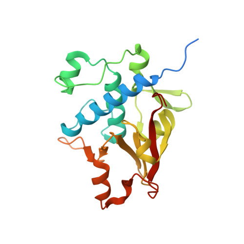
MagC is a NplC/P60-like member of the alpha-2-macroglobulin Mag complex of Pseudomonas aeruginosa that interacts with peptidoglycan.

Zouhir, S.Contreras-Martel, C.Maragno Trindade, D.Attree, I.Dessen, A.Macheboeuf, P.

(2021) FEBS Lett 595: 2034-2046

  • DOI: https://doi.org/10.1002/1873-3468.14148
  • Primary Citation of Related Structures:  
    7BK8

  • PubMed Abstract: 

    Bacterial α-2 macroglobulins (A2Ms) structurally resemble the large spectrum protease inhibitors of the eukaryotic immune system. In Pseudomonas aeruginosa, MagD acts as an A2M and is expressed within a six-gene operon encoding the MagA-F proteins. In this work, we employ isothermal calorimetry (ITC), analytical ultracentrifugation (AUC), and X-ray crystallography to investigate the function of MagC and show that MagC associates with the macroglobulin complex and with the peptidoglycan (PG). However, the catalytic residues of MagC display an inactive conformation that could suggest that it binds to PG but does not degrade it. We hypothesize that MagC could serve as an anchor between the MagD macroglobulin and the PG and could provide stabilization and/or regulation for the entire complex.


  • Organizational Affiliation

    Brazilian Biosciences National Laboratory (LNBio), CNPEM, Campinas, Brazil.


Macromolecules
Find similar proteins by:  (by identity cutoff)  |  3D Structure
Entity ID: 1
MoleculeChains Sequence LengthOrganismDetailsImage
MagC212Pseudomonas aeruginosa PAO1Mutation(s): 0 
Gene Names: PA4490
UniProt
Find proteins for Q9HVT1 (Pseudomonas aeruginosa (strain ATCC 15692 / DSM 22644 / CIP 104116 / JCM 14847 / LMG 12228 / 1C / PRS 101 / PAO1))
Explore Q9HVT1 
Go to UniProtKB:  Q9HVT1
Entity Groups  
Sequence Clusters30% Identity50% Identity70% Identity90% Identity95% Identity100% Identity
UniProt GroupQ9HVT1
Sequence Annotations
Expand
  • Reference Sequence
Small Molecules
Ligands 1 Unique
IDChains Name / Formula / InChI Key2D Diagram3D Interactions
PO4
Query on PO4

Download Ideal Coordinates CCD File 
B [auth A]PHOSPHATE ION
O4 P
NBIIXXVUZAFLBC-UHFFFAOYSA-K
Experimental Data & Validation

Experimental Data

  • Method: X-RAY DIFFRACTION
  • Resolution: 1.74 Å
  • R-Value Free: 0.222 
  • R-Value Work: 0.193 
  • Space Group: H 3
Unit Cell:
Length ( Å )Angle ( ˚ )
a = 110.737α = 90
b = 110.737β = 90
c = 41.11γ = 120
Software Package:
Software NamePurpose
REFMACrefinement
XDSdata reduction
XSCALEdata scaling
Arcimboldophasing

Structure Validation

View Full Validation Report


Entry History & Funding Information

Deposition Data


Funding OrganizationLocationGrant Number
Sao Paulo Research Foundation (FAPESP)Brazil2011/52067-6
Sao Paulo Research Foundation (FAPESP)Brazil2017/12436-9
French Infrastructure for Integrated Structural Biology (FRISBI)FranceANR-10-INBS-0005-02

Revision History  (Full details and data files)

  • Version 1.0: 2021-07-21
    Type: Initial release
  • Version 1.1: 2021-08-18
    Changes: Database references