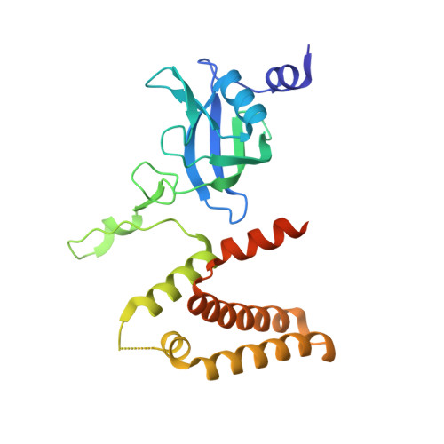
7BGM

Crystal structure of MtHISN2, a bifunctional enzyme from the histidine biosynthetic pathway


Experimental Data Snapshot

  • Method: X-RAY DIFFRACTION
  • Resolution: 1.60 Å
  • R-Value Free: 0.175 
  • R-Value Work: 0.143 
  • R-Value Observed: 0.144 

wwPDB Validation   3D Report Full Report


This is version 1.1 of the entry. See complete history


Literature

Structural and mechanistic insights into the bifunctional HISN2 enzyme catalyzing the second and third steps of histidine biosynthesis in plants.

Witek, W.Sliwiak, J.Ruszkowski, M.

(2021) Sci Rep 11: 9647-9647

  • DOI: https://doi.org/10.1038/s41598-021-88920-2
  • Primary Citation of Related Structures:  
    7BGM, 7BGN

  • PubMed Abstract: 

    The second and third steps of the histidine biosynthetic pathway (HBP) in plants are catalyzed by a bifunctional enzyme-HISN2. The enzyme consists of two distinct domains, active respectively as a phosphoribosyl-AMP cyclohydrolase (PRA-CH) and phosphoribosyl-ATP pyrophosphatase (PRA-PH). The domains are analogous to single-domain enzymes encoded by bacterial hisI and hisE genes, respectively. The calculated sequence similarity networks between HISN2 analogs from prokaryotes and eukaryotes suggest that the plant enzymes are closest relatives of those in the class of Deltaproteobacteria. In this work, we obtained crystal structures of HISN2 enzyme from Medicago truncatula (MtHISN2) and described its architecture and interactions with AMP. The AMP molecule bound to the PRA-PH domain shows positioning of the N1-phosphoribosyl relevant to catalysis. AMP bound to the PRA-CH domain mimics a part of the substrate, giving insights into the reaction mechanism. The latter interaction also arises as a possible second-tier regulatory mechanism of the HBP flux, as indicated by inhibition assays and isothermal titration calorimetry.


  • Organizational Affiliation

    Center for Biocrystallographic Research, Institute of Bioorganic Chemistry, Polish Academy of Sciences, Noskowskiego 12/14, 61-704, Poznan, Poland.


Macromolecules
Find similar proteins by:  (by identity cutoff)  |  3D Structure
Entity ID: 1
MoleculeChains Sequence LengthOrganismDetailsImage
Phosphoribosyl-AMP cyclohydrolase
A, B
238Medicago truncatulaMutation(s): 0 
Gene Names: 25498966MTR_7g084480MtrunA17_Chr7g0252441
EC: 3.5.4.19 (PDB Primary Data), 3.6.1.31 (PDB Primary Data), 3 (UniProt)
UniProt
Find proteins for A0A072U2X9 (Medicago truncatula)
Explore A0A072U2X9 
Go to UniProtKB:  A0A072U2X9
Entity Groups  
Sequence Clusters30% Identity50% Identity70% Identity90% Identity95% Identity100% Identity
UniProt GroupA0A072U2X9
Sequence Annotations
Expand
  • Reference Sequence
Small Molecules
Ligands 1 Unique
IDChains Name / Formula / InChI Key2D Diagram3D Interactions
ZN
Query on ZN

Download Ideal Coordinates CCD File 
C [auth A]
D [auth A]
E [auth A]
F [auth A]
G [auth B]
C [auth A],
D [auth A],
E [auth A],
F [auth A],
G [auth B],
H [auth B],
I [auth B],
J [auth B],
K [auth B]
ZINC ION
Zn
PTFCDOFLOPIGGS-UHFFFAOYSA-N
Experimental Data & Validation

Experimental Data

Unit Cell:
Length ( Å )Angle ( ˚ )
a = 172.422α = 90
b = 69.477β = 94.696
c = 52.13γ = 90
Software Package:
Software NamePurpose
PHENIXrefinement
XDSdata reduction
Cootmodel building
PHENIXmodel building
AutoSolphasing

Structure Validation

View Full Validation Report


Entry History & Funding Information

Deposition Data


Funding OrganizationLocationGrant Number
Polish National Science CentrePolandSONATA 2018/31/D/NZ1/03630

Revision History  (Full details and data files)

  • Version 1.0: 2021-05-19
    Type: Initial release
  • Version 1.1: 2024-05-15
    Changes: Data collection, Database references