7BF1

Ca2+-Calmodulin in complex with peptide from brain-type creatine kinase in extended 1:2 binding mode


Experimental Data Snapshot

  • Method: X-RAY DIFFRACTION
  • Resolution: 1.24 Å
  • R-Value Free: 0.186 
  • R-Value Work: 0.135 

wwPDB Validation   3D Report Full Report


This is version 1.1 of the entry. See complete history


Literature

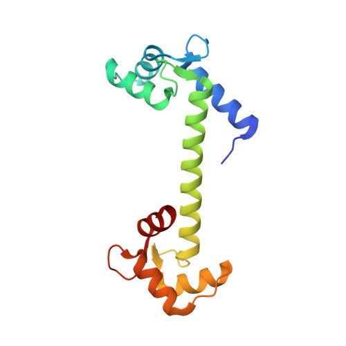
Calmodulin complexes with brain and muscle creatine kinase peptides.

Sprenger, J.Trifan, A.Patel, N.Vanderbeck, A.Bredfelt, J.Tajkhorshid, E.Rowlett, R.Lo Leggio, L.Akerfeldt, K.S.Linse, S.

(2021) Curr Res Struct Biol 3: 121-132

  • DOI: https://doi.org/10.1016/j.crstbi.2021.05.001
  • Primary Citation of Related Structures:  
    7BF1, 7BF2

  • PubMed Abstract: 

    Calmodulin (CaM) is a ubiquitous Ca 2+ sensing protein that binds to and modulates numerous target proteins and enzymes during cellular signaling processes. A large number of CaM-target complexes have been identified and structurally characterized, revealing a wide diversity of CaM-binding modes. A newly identified target is creatine kinase (CK), a central enzyme in cellular energy homeostasis. This study reports two high-resolution X-ray structures, determined to 1.24 ​Å and 1.43 ​Å resolution, of calmodulin in complex with peptides from human brain and muscle CK, respectively. Both complexes adopt a rare extended binding mode with an observed stoichiometry of 1:2 CaM:peptide, confirmed by isothermal titration calorimetry, suggesting that each CaM domain independently binds one CK peptide in a Ca 2+ -depended manner. While the overall binding mode is similar between the structures with muscle or brain-type CK peptides, the most significant difference is the opposite binding orientation of the peptides in the N-terminal domain. This may extrapolate into distinct binding modes and regulation of the full-length CK isoforms. The structural insights gained in this study strengthen the link between cellular energy homeostasis and Ca 2+ -mediated cell signaling and may shed light on ways by which cells can 'fine tune' their energy levels to match the spatial and temporal demands.


  • Organizational Affiliation

    Department of Biochemistry and Structural Biology, Chemical Center, PO Box 124, SE-221 00, Lund, Sweden.


Macromolecules
Find similar proteins by:  (by identity cutoff)  |  3D Structure
Entity ID: 1
MoleculeChains Sequence LengthOrganismDetailsImage
Calmodulin-1A [auth AAA]149Homo sapiensMutation(s): 0 
Gene Names: CALM1CALMCAMCAM1
UniProt & NIH Common Fund Data Resources
Find proteins for P0DP23 (Homo sapiens)
Explore P0DP23 
Go to UniProtKB:  P0DP23
PHAROS:  P0DP23
GTEx:  ENSG00000198668 
Entity Groups  
Sequence Clusters30% Identity50% Identity70% Identity90% Identity95% Identity100% Identity
UniProt GroupP0DP23
Sequence Annotations
Expand
  • Reference Sequence

Find similar proteins by:  Sequence   |   3D Structure  

Entity ID: 2
MoleculeChains Sequence LengthOrganismDetailsImage
Creatine kinase B-typeB [auth CCC],
C [auth DDD]
18Homo sapiensMutation(s): 0 
EC: 2.7.3.2
UniProt & NIH Common Fund Data Resources
Find proteins for P12277 (Homo sapiens)
Explore P12277 
Go to UniProtKB:  P12277
PHAROS:  P12277
GTEx:  ENSG00000166165 
Entity Groups  
Sequence Clusters30% Identity50% Identity70% Identity90% Identity95% Identity100% Identity
UniProt GroupP12277
Sequence Annotations
Expand
  • Reference Sequence
Experimental Data & Validation

Experimental Data

  • Method: X-RAY DIFFRACTION
  • Resolution: 1.24 Å
  • R-Value Free: 0.186 
  • R-Value Work: 0.135 
  • Space Group: P 1 21 1
Unit Cell:
Length ( Å )Angle ( ˚ )
a = 24.411α = 90
b = 59.534β = 98.343
c = 52.477γ = 90
Software Package:
Software NamePurpose
REFMACrefinement
XDSdata reduction
Aimlessdata scaling
MOLREPphasing

Structure Validation

View Full Validation Report


Entry History 

Deposition Data

Revision History  (Full details and data files)

  • Version 1.0: 2021-07-21
    Type: Initial release
  • Version 1.1: 2024-01-31
    Changes: Data collection, Database references, Derived calculations, Refinement description