7BBQ

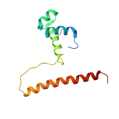
Crystal structure of the HTH DNA binding protein ArdK from R388 plasmid. Apo form.


Experimental Data Snapshot

  • Method: X-RAY DIFFRACTION
  • Resolution: 3.00 Å
  • R-Value Free: 0.271 
  • R-Value Work: 0.246 
  • R-Value Observed: 0.248 

wwPDB Validation   3D Report Full Report


This is version 1.2 of the entry. See complete history


Literature

Structural basis of direct and inverted DNA sequence repeat recognition by helix-turn-helix transcription factors.

Fernandez-Lopez, R.Ruiz, R.Del Campo, I.Gonzalez-Montes, L.Boer, D.R.de la Cruz, F.Moncalian, G.

(2022) Nucleic Acids Res 50: 11938-11947

  • DOI: https://doi.org/10.1093/nar/gkac1024
  • Primary Citation of Related Structures:  
    7BBQ, 7BCA, 7BCB

  • PubMed Abstract: 

    Some transcription factors bind DNA motifs containing direct or inverted sequence repeats. Preference for each of these DNA topologies is dictated by structural constraints. Most prokaryotic regulators form symmetric oligomers, which require operators with a dyad structure. Binding to direct repeats requires breaking the internal symmetry, a property restricted to a few regulators, most of them from the AraC family. The KorA family of transcriptional repressors, involved in plasmid propagation and stability, includes members that form symmetric dimers and recognize inverted repeats. Our structural analyses show that ArdK, a member of this family, can form a symmetric dimer similar to that observed for KorA, yet it binds direct sequence repeats as a non-symmetric dimer. This is possible by the 180° rotation of one of the helix-turn-helix domains. We then probed and confirmed that ArdK shows affinity for an inverted repeat, which, surprisingly, is also recognized by a non-symmetrical dimer. Our results indicate that structural flexibility at different positions in the dimerization interface constrains transcription factors to bind DNA sequences with one of these two alternative DNA topologies.


  • Organizational Affiliation

    Departamento de Biología Molecular, Universidad de Cantabria and Instituto de Biomedicina y Biotecnología de Cantabria (IBBTEC), CSIC-Universidad de Cantabria, 39011, Santander, Spain.


Macromolecules
Find similar proteins by:  (by identity cutoff)  |  3D Structure
Entity ID: 1
MoleculeChains Sequence LengthOrganismDetailsImage
KORA domain-containing protein110Escherichia coli K-12Mutation(s): 1 
UniProt
Find proteins for Q6I6B7 (Escherichia coli)
Explore Q6I6B7 
Go to UniProtKB:  Q6I6B7
Entity Groups  
Sequence Clusters30% Identity50% Identity70% Identity90% Identity95% Identity100% Identity
UniProt GroupQ6I6B7
Sequence Annotations
Expand
  • Reference Sequence
Small Molecules
Modified Residues  1 Unique
IDChains TypeFormula2D DiagramParent
MSE
Query on MSE
A
L-PEPTIDE LINKINGC5 H11 N O2 SeMET
Experimental Data & Validation

Experimental Data

  • Method: X-RAY DIFFRACTION
  • Resolution: 3.00 Å
  • R-Value Free: 0.271 
  • R-Value Work: 0.246 
  • R-Value Observed: 0.248 
  • Space Group: P 41 21 2
Unit Cell:
Length ( Å )Angle ( ˚ )
a = 43.1α = 90
b = 43.1β = 90
c = 190.89γ = 90
Software Package:
Software NamePurpose
PHENIXrefinement
PDB_EXTRACTdata extraction
SCALAdata scaling
PHENIXphasing
MOSFLMdata reduction
Cootmodel building

Structure Validation

View Full Validation Report


Entry History & Funding Information

Deposition Data


Funding OrganizationLocationGrant Number
Spanish Ministry of Economy and CompetitivenessSpainBFU2014-55534-C2-2-P
Spanish Ministry of Science, Innovation, and UniversitiesSpainPGC2018-093885-B-I00

Revision History  (Full details and data files)

  • Version 1.0: 2022-07-13
    Type: Initial release
  • Version 1.1: 2022-11-23
    Changes: Database references
  • Version 1.2: 2022-12-14
    Changes: Database references