7B8Z | pdb_00007b8z

Notum-Fragment 277


Experimental Data Snapshot

  • Method: X-RAY DIFFRACTION
  • Resolution: 1.73 Å
  • R-Value Free: 
    0.243 (Depositor), 0.247 (DCC) 
  • R-Value Work: 
    0.227 (Depositor), 0.229 (DCC) 
  • R-Value Observed: 
    0.227 (Depositor) 

Starting Model: experimental
View more details

wwPDB Validation 3D Report Full Report

Validation slider image for 7B8Z

Ligand Structure Quality Assessment 


This is version 1.3 of the entry. See complete history

Literature

Structural Analysis and Development of Notum Fragment Screening Hits.

Zhao, Y.Mahy, W.Willis, N.J.Woodward, H.L.Steadman, D.Bayle, E.D.Atkinson, B.N.Sipthorp, J.Vecchia, L.Ruza, R.R.Harlos, K.Jeganathan, F.Constantinou, S.Costa, A.Kjaer, S.Bictash, M.Salinas, P.C.Whiting, P.Vincent, J.P.Fish, P.V.Jones, E.Y.

(2022) ACS Chem Neurosci 13: 2060-2077

  • DOI: https://doi.org/10.1021/acschemneuro.2c00325
  • Primary Citation Related Structures: 
    7B4X, 7B84, 7B86, 7B87, 7B89, 7B8C, 7B8D, 7B8F, 7B8G, 7B8J, 7B8K, 7B8L, 7B8M, 7B8N, 7B8O, 7B8U, 7B8X, 7B8Y, 7B8Z, 7B98, 7B99, 7B9D, 7B9I, 7B9N, 7B9U, 7BA1, 7BAC, 7BAP, 7BC8, 7BC9, 7BCC, 7BCD, 7BCE, 7BCF, 7BCH, 7BCI, 7BCK, 7BCL, 7BD2, 7BD3, 7BD4, 7BD5, 7BD6, 7BD8, 7BD9, 7BDA, 7BDB, 7BDC, 7BDD, 7BDF, ... Search all related entries

  • PubMed Abstract: 

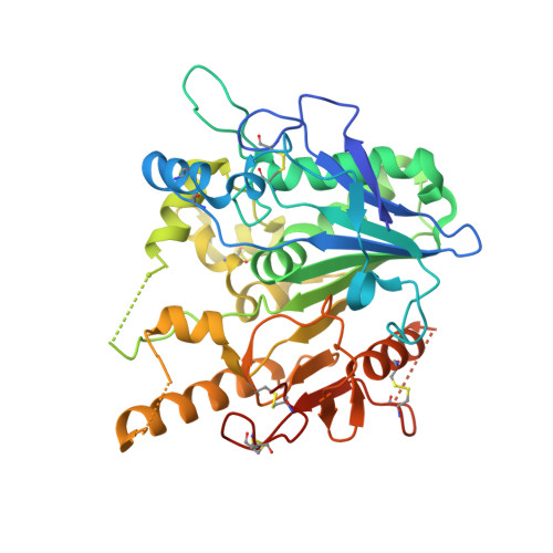
    The Wnt signaling suppressor Notum is a promising target for osteoporosis, Alzheimer's disease, and colorectal cancers. To develop novel Notum inhibitors, we used an X-ray crystallographic fragment screen with the Diamond-SGC Poised Library (DSPL) and identified 59 fragment hits from the analysis of 768 data sets. Fifty-eight of the hits were found bound at the enzyme catalytic pocket with potencies ranging from 0.5 to >1000 μM. Analysis of the fragments' diverse binding modes, enzymatic inhibitory activities, and chemical properties led to the selection of six hits for optimization, and five of these resulted in improved Notum inhibitory potencies. One hit, 1 - phenyl-1,2,3-triazole 7 , and its related cluster members, have shown promising lead-like properties. These became the focus of our fragment development activities, resulting in compound 7d with IC 50 0.0067 μM. The large number of Notum fragment structures and their initial optimization provided an important basis for further Notum inhibitor development.


  • Organizational Affiliation
    • Division of Structural Biology, Wellcome Centre for Human Genetics, University of Oxford, The Henry Wellcome Building for Genomic Medicine, Roosevelt Drive, Oxford OX3 7BN, U.K.

Macromolecule Content 

  • Total Structure Weight: 44.78 kDa 
  • Atom Count: 2,826 
  • Modeled Residue Count: 342 
  • Deposited Residue Count: 383 
  • Unique protein chains: 1

Macromolecules

Find similar proteins by:|  3D Structure
Entity ID: 1
MoleculeChains  Sequence LengthOrganismDetailsImage
Palmitoleoyl-protein carboxylesterase NOTUM383Homo sapiensMutation(s): 1 
Gene Names: NOTUMOK/SW-CL.30
EC: 3.1.1.98
UniProt & NIH Common Fund Data Resources
Find proteins for Q6P988 (Homo sapiens)
Explore Q6P988 
Go to UniProtKB:  Q6P988
PHAROS:  Q6P988
GTEx:  ENSG00000185269 
Entity Groups
Sequence Clusters30% Identity50% Identity70% Identity90% Identity95% Identity100% Identity
UniProt GroupQ6P988
Glycosylation
Glycosylation Sites: 1Go to GlyGen: Q6P988-1
Sequence Annotations
Expand
Reference Sequence

Small Molecules

Ligands 4 Unique
IDChains Name / Formula / InChI Key2D Diagram3D Interactions
A6Z
(Subject of Investigation/LOI)

Query on A6Z


Download:Ideal Coordinates CCD File
K [auth A]2-[4-(3-chlorophenyl)piperazin-1-ium-1-yl]ethanenitrile
C12 H15 Cl N3
BIGOTGAMVFQWPQ-UHFFFAOYSA-O
NAG

Query on NAG


Download:Ideal Coordinates CCD File
C [auth A]2-acetamido-2-deoxy-beta-D-glucopyranose
C8 H15 N O6
OVRNDRQMDRJTHS-FMDGEEDCSA-N
SO4

Query on SO4


Download:Ideal Coordinates CCD File
B [auth A]
E [auth A]
F [auth A]
G [auth A]
H [auth A]
B [auth A],
E [auth A],
F [auth A],
G [auth A],
H [auth A],
I [auth A],
J [auth A]
SULFATE ION
O4 S
QAOWNCQODCNURD-UHFFFAOYSA-L
DMS

Query on DMS


Download:Ideal Coordinates CCD File
D [auth A]DIMETHYL SULFOXIDE
C2 H6 O S
IAZDPXIOMUYVGZ-UHFFFAOYSA-N

Experimental Data & Validation

Experimental Data

  • Method: X-RAY DIFFRACTION
  • Resolution: 1.73 Å
  • R-Value Free:  0.243 (Depositor), 0.247 (DCC) 
  • R-Value Work:  0.227 (Depositor), 0.229 (DCC) 
  • R-Value Observed: 0.227 (Depositor) 
Space Group: P 21 21 21
Unit Cell:
Length ( Å )Angle ( ˚ )
a = 59.457α = 90
b = 73.598β = 90
c = 78.758γ = 90
Software Package:
Software NamePurpose
xia2data scaling
PHENIXrefinement
PDB_EXTRACTdata extraction
xia2data reduction
MOLREPphasing

Structure Validation

View Full Validation Report


Ligand Structure Quality Assessment 


Entry History 

& Funding Information

Deposition Data


Funding OrganizationLocationGrant Number
Cancer Research UKUnited KingdomC375/A17721

Revision History  (Full details and data files)

  • Version 1.0: 2022-01-12
    Type: Initial release
  • Version 1.1: 2022-07-27
    Changes: Database references
  • Version 1.2: 2024-01-31
    Changes: Data collection, Refinement description
  • Version 1.3: 2024-10-16
    Changes: Structure summary