7B1H

Monoclinic P21 Structure of Human Mad1 C-terminal Domain in Complex with Phosphorylated Bub1 CD1 Domain


Experimental Data Snapshot

  • Method: X-RAY DIFFRACTION
  • Resolution: 2.40 Å
  • R-Value Free: 0.281 
  • R-Value Work: 0.231 
  • R-Value Observed: 0.234 

wwPDB Validation   3D Report Full Report


This is version 1.3 of the entry. See complete history


Literature

Molecular mechanism of Mad1 kinetochore targeting by phosphorylated Bub1.

Fischer, E.S.Yu, C.W.H.Bellini, D.McLaughlin, S.H.Orr, C.M.Wagner, A.Freund, S.M.V.Barford, D.

(2021) EMBO Rep 22: e52242-e52242

  • DOI: https://doi.org/10.15252/embr.202052242
  • Primary Citation of Related Structures:  
    7B1F, 7B1H, 7B1J

  • PubMed Abstract: 

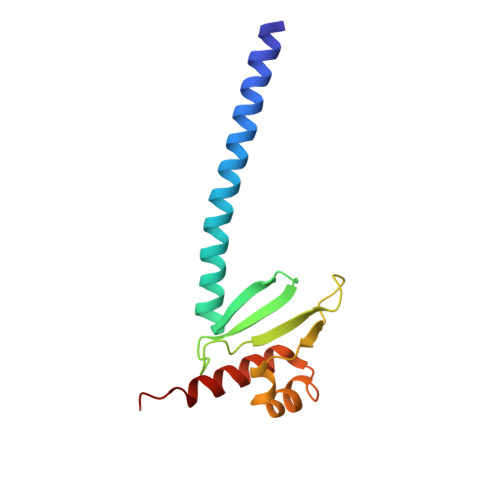
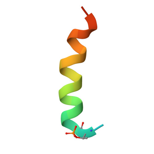
    During metaphase, in response to improper kinetochore-microtubule attachments, the spindle assembly checkpoint (SAC) activates the mitotic checkpoint complex (MCC), an inhibitor of the anaphase-promoting complex/cyclosome (APC/C). This process is orchestrated by the kinase Mps1, which initiates the assembly of the MCC onto kinetochores through a sequential phosphorylation-dependent signalling cascade. The Mad1-Mad2 complex, which is required to catalyse MCC formation, is targeted to kinetochores through a direct interaction with the phosphorylated conserved domain 1 (CD1) of Bub1. Here, we present the crystal structure of the C-terminal domain of Mad1 (Mad1 CTD ) bound to two phosphorylated Bub1 CD1 peptides at 1.75 Å resolution. This interaction is mediated by phosphorylated Bub1 Thr461, which not only directly interacts with Arg617 of the Mad1 RLK (Arg-Leu-Lys) motif, but also directly acts as an N-terminal cap to the CD1 α-helix dipole. Surprisingly, only one Bub1 CD1 peptide binds to the Mad1 homodimer in solution. We suggest that this stoichiometry is due to inherent asymmetry in the coiled-coil of Mad1 CTD and has implications for how the Mad1-Bub1 complex at kinetochores promotes efficient MCC assembly.


  • Organizational Affiliation

    MRC Laboratory of Molecular Biology, Cambridge, UK.


Macromolecules
Find similar proteins by:  (by identity cutoff)  |  3D Structure
Entity ID: 1
MoleculeChains Sequence LengthOrganismDetailsImage
Mitotic spindle assembly checkpoint protein MAD1A [auth B],
B [auth A],
E,
F
122Homo sapiensMutation(s): 0 
Gene Names: MAD1L1MAD1TXBP181
UniProt & NIH Common Fund Data Resources
Find proteins for Q9Y6D9 (Homo sapiens)
Explore Q9Y6D9 
Go to UniProtKB:  Q9Y6D9
PHAROS:  Q9Y6D9
GTEx:  ENSG00000002822 
Entity Groups  
Sequence Clusters30% Identity50% Identity70% Identity90% Identity95% Identity100% Identity
UniProt GroupQ9Y6D9
Sequence Annotations
Expand
  • Reference Sequence
Find similar proteins by:  (by identity cutoff)  |  3D Structure
Entity ID: 2
MoleculeChains Sequence LengthOrganismDetailsImage
Mitotic checkpoint serine/threonine-protein kinase BUB1
C, D, G, H
26Homo sapiensMutation(s): 0 
EC: 2.7.11.1
UniProt & NIH Common Fund Data Resources
Find proteins for O43683 (Homo sapiens)
Explore O43683 
Go to UniProtKB:  O43683
PHAROS:  O43683
GTEx:  ENSG00000169679 
Entity Groups  
Sequence Clusters30% Identity50% Identity70% Identity90% Identity95% Identity100% Identity
UniProt GroupO43683
Sequence Annotations
Expand
  • Reference Sequence
Small Molecules
Modified Residues  2 Unique
IDChains TypeFormula2D DiagramParent
SEP
Query on SEP
C, D, G, H
L-PEPTIDE LINKINGC3 H8 N O6 PSER
TPO
Query on TPO
C, D, G, H
L-PEPTIDE LINKINGC4 H10 N O6 PTHR
Experimental Data & Validation

Experimental Data

  • Method: X-RAY DIFFRACTION
  • Resolution: 2.40 Å
  • R-Value Free: 0.281 
  • R-Value Work: 0.231 
  • R-Value Observed: 0.234 
  • Space Group: P 1 21 1
Unit Cell:
Length ( Å )Angle ( ˚ )
a = 34.63α = 90
b = 132.34β = 93.34
c = 82.75γ = 90
Software Package:
Software NamePurpose
PHENIXrefinement
xia2data reduction
XDSdata scaling
PHASERphasing

Structure Validation

View Full Validation Report


Entry History & Funding Information

Deposition Data


Funding OrganizationLocationGrant Number
Medical Research Council (MRC, United Kingdom)United KingdomMC_UP_1201/6
Cancer Research UKUnited KingdomC576/A14109

Revision History  (Full details and data files)

  • Version 1.0: 2021-03-17
    Type: Initial release
  • Version 1.1: 2021-06-02
    Changes: Database references
  • Version 1.2: 2021-07-14
    Changes: Database references
  • Version 1.3: 2024-01-31
    Changes: Data collection, Database references, Refinement description