Yeast Ssd1 is a non-enzymatic member of the RNase II family with an alternative RNA recognition site.
Bayne, R.A., Jayachandran, U., Kasprowicz, A., Bresson, S., Tollervey, D., Wallace, E.W.J., Cook, A.G.(2022) Nucleic Acids Res 50: 2923-2937
- PubMed: 34302485 Search on PubMedSearch on PubMed Central
- DOI: https://doi.org/10.1093/nar/gkab615
- Primary Citation Related Structures:
7AM1 - PubMed Abstract:
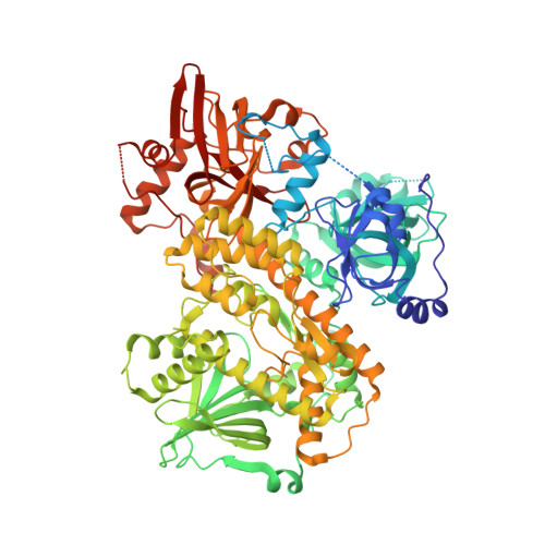
Ssd1, a conserved fungal RNA-binding protein, is important in stress responses, cell division and virulence. Ssd1 is closely related to Dis3L2 of the RNase II family of nucleases, but lacks catalytic activity and likely suppresses translation of bound mRNAs. Previous studies identified RNA motifs enriched in Ssd1-associated transcripts, yet the sequence requirements for Ssd1 binding are not defined. Here, we identify precise binding sites of Ssd1 on RNA using in vivo cross-linking and cDNA analysis. These sites are enriched in 5' untranslated regions of a subset of mRNAs encoding cell wall proteins. We identified a conserved bipartite motif that binds Ssd1 with high affinity in vitro. Active RNase II enzymes have a characteristic, internal RNA binding path; the Ssd1 crystal structure at 1.9 Å resolution shows that remnants of regulatory sequences block this path. Instead, RNA binding activity has relocated to a conserved patch on the surface of the protein. Structure-guided mutations of this surface prevent Ssd1 from binding RNA in vitro and phenocopy Ssd1 deletion in vivo. These studies provide a new framework for understanding the function of a pleiotropic post-transcriptional regulator of gene expression and give insights into the evolution of regulatory and binding elements in the RNase II family.
- Institute of Cell Biology and SynthSys, School of Biological Sciences, University of Edinburgh, Edinburgh EH9 3BF, UK.
Organizational Affiliation: