The conserved C2 phospholipid-binding domain in Delta contributes to robust Notch signalling.
Martins, T., Meng, Y., Korona, B., Suckling, R., Johnson, S., Handford, P.A., Lea, S.M., Bray, S.J.(2021) EMBO Rep 22: e52729-e52729
- PubMed: 34347930 Search on PubMedSearch on PubMed Central
- DOI: https://doi.org/10.15252/embr.202152729
- Primary Citation Related Structures:
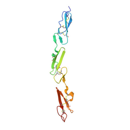
7ALJ, 7ALK, 7ALT - PubMed Abstract:
Accurate Notch signalling is critical for development and homeostasis. Fine-tuning of Notch-ligand interactions has substantial impact on signalling outputs. Recent structural studies have identified a conserved N-terminal C2 domain in human Notch ligands which confers phospholipid binding in vitro. Here, we show that Drosophila ligands Delta and Serrate adopt the same C2 domain structure with analogous variations in the loop regions, including the so-called β1-2 loop that is involved in phospholipid binding. Mutations in the β1-2 loop of the Delta C2 domain retain Notch binding but have impaired ability to interact with phospholipids in vitro. To investigate its role in vivo, we deleted five residues within the β1-2 loop of endogenous Delta. Strikingly, this change compromises ligand function. The modified Delta enhances phenotypes produced by Delta loss-of-function alleles and suppresses that of Notch alleles. As the modified protein is present on the cell surface in normal amounts, these results argue that C2 domain phospholipid binding is necessary for robust signalling in vivo fine-tuning the balance of trans and cis ligand-receptor interactions.
- Department of Physiology Development and Neuroscience, University of Cambridge, Cambridge, UK.
Organizational Affiliation: