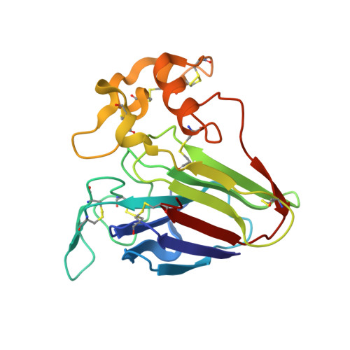
Versatile microporous polymer-based supports for serial macromolecular crystallography.
Martiel, I., Beale, J.H., Karpik, A., Huang, C.Y., Vera, L., Olieric, N., Wranik, M., Tsai, C.J., Muhle, J., Aurelius, O., John, J., Hogbom, M., Wang, M., Marsh, M., Padeste, C.(2021) Acta Crystallogr D Struct Biol 77: 1153-1167
- PubMed: 34473086
- DOI: https://doi.org/10.1107/S2059798321007324
- Primary Citation of Related Structures:
7AC2, 7AC3, 7AC4, 7AC5, 7AC6, 7AI8, 7AI9 - PubMed Abstract:
Serial data collection has emerged as a major tool for data collection at state-of-the-art light sources, such as microfocus beamlines at synchrotrons and X-ray free-electron lasers. Challenging targets, characterized by small crystal sizes, weak diffraction and stringent dose limits, benefit most from these methods. Here, the use of a thin support made of a polymer-based membrane for performing serial data collection or screening experiments is demonstrated. It is shown that these supports are suitable for a wide range of protein crystals suspended in liquids. The supports have also proved to be applicable to challenging cases such as membrane proteins growing in the sponge phase. The sample-deposition method is simple and robust, as well as flexible and adaptable to a variety of cases. It results in an optimally thin specimen providing low background while maintaining minute amounts of mother liquor around the crystals. The 2 × 2 mm area enables the deposition of up to several microlitres of liquid. Imaging and visualization of the crystals are straightforward on the highly transparent membrane. Thanks to their affordable fabrication, these supports have the potential to become an attractive option for serial experiments at synchrotrons and free-electron lasers.
- Paul Scherrer Institute, Forschungsstrasse 111, 5232 Villigen, Switzerland.
Organizational Affiliation:


