Structural insights into a novel family of integral membrane siderophore reductases.
Josts, I., Veith, K., Normant, V., Schalk, I.J., Tidow, H.(2021) Proc Natl Acad Sci U S A 118
- PubMed: 34417315 Search on PubMedSearch on PubMed Central
- DOI: https://doi.org/10.1073/pnas.2101952118
- Primary Citation Related Structures:
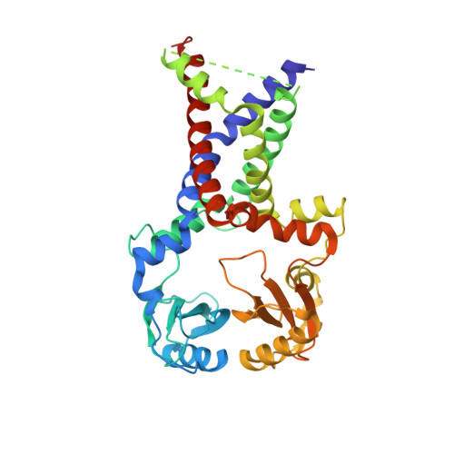
7ABW - PubMed Abstract:
Gram-negative bacteria take up the essential ion Fe 3+ as ferric-siderophore complexes through their outer membrane using TonB-dependent transporters. However, the subsequent route through the inner membrane differs across many bacterial species and siderophore chemistries and is not understood in detail. Here, we report the crystal structure of the inner membrane protein FoxB (from Pseudomonas aeruginosa ) that is involved in Fe-siderophore uptake. The structure revealed a fold with two tightly bound heme molecules. In combination with in vitro reduction assays and in vivo iron uptake studies, these results establish FoxB as an inner membrane reductase involved in the release of iron from ferrioxamine during Fe-siderophore uptake.
- The Hamburg Advanced Research Center for Bioorganic Chemistry (HARBOR), 22761 Hamburg, Germany; tidow@chemie.uni-hamburg.de josts@chemie.uni-hamburg.de.
Organizational Affiliation: