7AAT | pdb_00007aat

X-RAY STRUCTURE REFINEMENT AND COMPARISON OF THREE FORMS OF MITOCHONDRIAL ASPARTATE AMINOTRANSFERASE


Experimental Data Snapshot

  • Method: X-RAY DIFFRACTION
  • Resolution: 1.90 Å
  • R-Value Observed: 
    0.166 (Depositor) 

wwPDB Validation 3D Report Full Report

Validation slider image for 7AAT

This is version 1.4 of the entry. See complete history

Literature

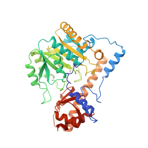
X-ray structure refinement and comparison of three forms of mitochondrial aspartate aminotransferase.

McPhalen, C.A.Vincent, M.G.Jansonius, J.N.

(1992) J Mol Biology 225: 495-517

  • DOI: https://doi.org/10.1016/0022-2836(92)90935-d
  • Primary Citation Related Structures: 
    7AAT, 8AAT, 9AAT

  • PubMed Abstract: 

    The X-ray crystal structures of three forms of the enzyme aspartate aminotransferase (EC 2.6.1.1) from chicken heart mitochondria have been refined by least-squares methods: holoenzyme with the co-factor pyridoxal-5'-phosphate bound at pH 7.5 (1.9 A resolution), holoenzyme with pyridoxal-5'-phosphate bound at pH 5.1 (2.3 A resolution) and holoenzyme with the co-factor pyridoxamine-5'-phosphate bound at pH 7.5 (2.2 A resolution). The crystallographic agreement factors [formula: see text] for the structures are 0.166, 0.130 and 0.131, respectively, for all data in the resolution range from 10.0 A to the limit of diffraction for each structure. The secondary, super-secondary and domain structures of the pyridoxal-phosphate holoenzyme at pH 7.5 are described in detail. The surface area of the interface between the monomer subunits of this dimeric alpha 2 protein is unusually large, indicating a very stable dimer. This is consistent with biochemical data. Both subunit and domain interfaces are relatively smooth compared with other proteins. The interactions of the protein with its co-factor are described and compared among the three structures. Observed changes in co-factor conformation may be related to spectral changes and the energetics of the catalytic reaction. Small but significant adjustments of the protein to changes in co-factor conformation are seen. These adjustments may be accommodated by small rigid-body shifts of secondary structural elements, and by packing defects in the protein core.


  • Organizational Affiliation
    • Department of Structural Biology, University of Basel, Switzerland.

Macromolecule Content 

  • Total Structure Weight: 90.48 kDa 
  • Atom Count: 7,022 
  • Modeled Residue Count: 802 
  • Deposited Residue Count: 802 
  • Unique protein chains: 1

Macromolecules

Find similar proteins by:|  3D Structure
Entity ID: 1
MoleculeChains  Sequence LengthOrganismDetailsImage
ASPARTATE AMINOTRANSFERASE
A, B
401Gallus gallusMutation(s): 0 
EC: 2.6.1.1 (PDB Primary Data), 2.6.1.7 (UniProt)
UniProt
Find proteins for P00508 (Gallus gallus)
Explore P00508 
Go to UniProtKB:  P00508
Entity Groups
Sequence Clusters30% Identity50% Identity70% Identity90% Identity95% Identity100% Identity
UniProt GroupP00508
Sequence Annotations
Expand
Reference Sequence

Small Molecules

Ligands 1 Unique
IDChains Name / Formula / InChI Key2D Diagram3D Interactions
PLP

Query on PLP


Download:Ideal Coordinates CCD File
C [auth A],
D [auth B]
PYRIDOXAL-5'-PHOSPHATE
C8 H10 N O6 P
NGVDGCNFYWLIFO-UHFFFAOYSA-N

Experimental Data & Validation

Experimental Data

  • Method: X-RAY DIFFRACTION
  • Resolution: 1.90 Å
  • R-Value Observed: 0.166 (Depositor) 
Space Group: P 1
Unit Cell:
Length ( Å )Angle ( ˚ )
a = 55.6α = 85.2
b = 58.7β = 109.2
c = 75.9γ = 115.6
Software Package:
Software NamePurpose
PROLSQrefinement

Structure Validation

View Full Validation Report


Entry History 

Deposition Data

Revision History  (Full details and data files)

  • Version 1.0: 1993-10-31
    Type: Initial release
  • Version 1.1: 2008-03-03
    Changes: Version format compliance
  • Version 1.2: 2011-07-13
    Changes: Non-polymer description, Version format compliance
  • Version 1.3: 2017-11-29
    Changes: Derived calculations, Other
  • Version 1.4: 2025-03-26
    Changes: Data collection, Database references, Derived calculations, Structure summary