Structural studies of the shortest extended synaptotagmin with only two C2 domains from Trypanosoma brucei .
Stepinac, E., Landrein, N., Skwarzynska, D., Wojcik, P., Lesigang, J., Lucic, I., He, C.Y., Bonhivers, M., Robinson, D.R., Dong, G.(2021) iScience 24: 102422-102422
- PubMed: 33997700 Search on PubMedSearch on PubMed Central
- DOI: https://doi.org/10.1016/j.isci.2021.102422
- Primary Citation Related Structures:
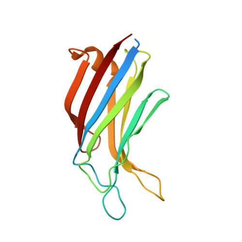
7A1R - PubMed Abstract:
Extended synaptotagmins (E-Syts) localize at membrane contact sites between the endoplasmic reticulum (ER) and the plasma membrane to mediate inter-membrane lipid transfer and control plasma membrane lipid homeostasis. All known E-Syts contain an N-terminal transmembrane (TM) hairpin, a central synaptotagmin-like mitochondrial lipid-binding protein (SMP) domain, and three or five C2 domains at their C termini. Here we report an uncharacterized E-Syt from the protist parasite Trypanosoma brucei , namely, TbE-Syt. TbE-Syt contains only two C2 domains (C2A and C2B), making it the shortest E-Syt known by now. We determined a 1.5-Å-resolution crystal structure of TbE-Syt-C2B and revealed that it binds lipids via both Ca 2+ - and PI(4,5)P 2 -dependent means. In contrast, TbE-Syt-C2A lacks the Ca 2+ -binding site but may still interact with lipids via a basic surface patch. Our studies suggest a mechanism for how TbE-Syt tethers the ER membrane tightly to the plasma membrane to transfer lipids between the two organelles.
- Max Perutz Labs, Vienna Biocenter, Center for Medical Biochemistry, Medical University of Vienna, 1030 Vienna, Austria.
Organizational Affiliation: