6YSA

Crystal structure of Arabidopsis thaliana legumain isoform beta in zymogen state


Experimental Data Snapshot

  • Method: X-RAY DIFFRACTION
  • Resolution: 2.01 Å
  • R-Value Free: 0.219 
  • R-Value Work: 0.209 
  • R-Value Observed: 0.209 

wwPDB Validation   3D Report Full Report


This is version 2.1 of the entry. See complete history


Literature

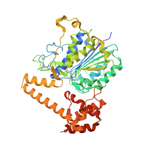
Structural and functional studies ofArabidopsis thalianalegumain beta reveal isoform specific mechanisms of activation and substrate recognition.

Dall, E.Zauner, F.B.Soh, W.T.Demir, F.Dahms, S.O.Cabrele, C.Huesgen, P.F.Brandstetter, H.

(2020) J Biol Chem 295: 13047-13064

  • DOI: https://doi.org/10.1074/jbc.RA120.014478
  • Primary Citation of Related Structures:  
    6YSA

  • PubMed Abstract: 

    The vacuolar cysteine protease legumain plays important functions in seed maturation and plant programmed cell death. Because of their dual protease and ligase activity, plant legumains have become of particular biotechnological interest, e.g. for the synthesis of cyclic peptides for drug design or for protein engineering. However, the molecular mechanisms behind their dual protease and ligase activities are still poorly understood, limiting their applications. Here, we present the crystal structure of Arabidopsis thaliana legumain isoform β (AtLEGβ) in its zymogen state. Combining structural and biochemical experiments, we show for the first time that plant legumains encode distinct, isoform-specific activation mechanisms. Whereas the autocatalytic activation of isoform γ (AtLEGγ) is controlled by the latency-conferring dimer state, the activation of the monomeric AtLEGβ is concentration independent. Additionally, in AtLEGβ the plant-characteristic two-chain intermediate state is stabilized by hydrophobic rather than ionic interactions, as in AtLEGγ, resulting in significantly different pH stability profiles. The crystal structure of AtLEGβ revealed unrestricted nonprime substrate binding pockets, consistent with the broad substrate specificity, as determined by degradomic assays. Further to its protease activity, we show that AtLEGβ exhibits a true peptide ligase activity. Whereas cleavage-dependent transpeptidase activity has been reported for other plant legumains, AtLEGβ is the first example of a plant legumain capable of linking free termini. The discovery of these isoform-specific differences will allow us to identify and rationally design efficient ligases with application in biotechnology and drug development.


  • Organizational Affiliation

    Department of Biosciences, University of Salzburg, Salzburg, Austria. Electronic address: elfriede.dall@sbg.ac.at.


Macromolecules
Find similar proteins by:  (by identity cutoff)  |  3D Structure
Entity ID: 1
MoleculeChains Sequence LengthOrganismDetailsImage
Vacuolar-processing enzyme beta-isozyme
A, B, C, D, E
A, B, C, D, E, F, G, H, I, J, K, L
455Arabidopsis thalianaMutation(s): 1 
Gene Names: bVPEAt1g62710F23N19.7
EC: 3.4.22.34
UniProt
Find proteins for Q39044 (Arabidopsis thaliana)
Explore Q39044 
Go to UniProtKB:  Q39044
Entity Groups  
Sequence Clusters30% Identity50% Identity70% Identity90% Identity95% Identity100% Identity
UniProt GroupQ39044
Sequence Annotations
Expand
  • Reference Sequence
Oligosaccharides

Help

Entity ID: 2
MoleculeChains Length2D Diagram Glycosylation3D Interactions
2-acetamido-2-deoxy-beta-D-glucopyranose-(1-4)-2-acetamido-2-deoxy-beta-D-glucopyranose
M, N, O, P, Q
M, N, O, P, Q, R, S, T, U, V, W, X
2N-Glycosylation
Glycosylation Resources
GlyTouCan:  G42666HT
GlyCosmos:  G42666HT
GlyGen:  G42666HT
Small Molecules
Ligands 2 Unique
IDChains Name / Formula / InChI Key2D Diagram3D Interactions
CIT
Query on CIT

Download Ideal Coordinates CCD File 
XA [auth E]CITRIC ACID
C6 H8 O7
KRKNYBCHXYNGOX-UHFFFAOYSA-N
SO4
Query on SO4

Download Ideal Coordinates CCD File 
AA [auth A]
AB [auth F]
AC [auth J]
BA [auth A]
BB [auth F]
AA [auth A],
AB [auth F],
AC [auth J],
BA [auth A],
BB [auth F],
BC [auth J],
CA [auth A],
CB [auth F],
CC [auth J],
DA [auth B],
DB [auth F],
DC [auth J],
EA [auth B],
EB [auth F],
EC [auth K],
FA [auth B],
FB [auth F],
FC [auth K],
GA [auth B],
GB [auth G],
GC [auth K],
HA [auth B],
HB [auth G],
HC [auth K],
IA [auth B],
IB [auth G],
IC [auth K],
JA [auth C],
JB [auth G],
JC [auth L],
KA [auth C],
KB [auth H],
KC [auth L],
LA [auth C],
LB [auth H],
LC [auth L],
MA [auth C],
MB [auth H],
MC [auth L],
NA [auth C],
NB [auth H],
OA [auth C],
OB [auth H],
PA [auth D],
PB [auth H],
QA [auth D],
QB [auth H],
RA [auth D],
RB [auth I],
SA [auth D],
SB [auth I],
TA [auth E],
TB [auth I],
UA [auth E],
UB [auth I],
VA [auth E],
VB [auth I],
WA [auth E],
WB [auth I],
XB [auth I],
Y [auth A],
YA [auth E],
YB [auth I],
Z [auth A],
ZA [auth F],
ZB [auth J]
SULFATE ION
O4 S
QAOWNCQODCNURD-UHFFFAOYSA-L
Modified Residues  1 Unique
IDChains TypeFormula2D DiagramParent
SNN
Query on SNN
A, B, C, D, E
A, B, C, D, E, F, G, H, I, J, K, L
L-PEPTIDE LINKINGC4 H6 N2 O2ASN
Experimental Data & Validation

Experimental Data

  • Method: X-RAY DIFFRACTION
  • Resolution: 2.01 Å
  • R-Value Free: 0.219 
  • R-Value Work: 0.209 
  • R-Value Observed: 0.209 
  • Space Group: P 41
Unit Cell:
Length ( Å )Angle ( ˚ )
a = 170.465α = 90
b = 170.465β = 90
c = 196.515γ = 90
Software Package:
Software NamePurpose
PHENIXrefinement
XDSdata reduction
XSCALEdata scaling
PHASERphasing

Structure Validation

View Full Validation Report


Entry History & Funding Information

Deposition Data


Funding OrganizationLocationGrant Number
Austrian Science FundAustriaW_01213

Revision History  (Full details and data files)

  • Version 1.0: 2020-07-29
    Type: Initial release
  • Version 1.1: 2020-08-12
    Changes: Database references
  • Version 1.2: 2020-09-23
    Changes: Database references
  • Version 2.0: 2023-11-15
    Changes: Advisory, Atomic model, Data collection, Database references, Derived calculations
  • Version 2.1: 2024-01-24
    Changes: Refinement description