


Substrate divergence in 7,8-diaminopelargonic acid synthesis: mutagenesis and computational studies of L-lysine dependent Bacillus subtilis BioA
Cramer, J., Kubota, C., Souza, S.A., Morris, J., Ng, H.L., Sun, R., Jarrett, J.T.To be published.
Experimental Data Snapshot
Starting Model: experimental
View more details
Entity ID: 1 | |||||
|---|---|---|---|---|---|
| Molecule | Chains | Sequence Length | Organism | Details | Image |
| Adenosylmethionine-8-amino-7-oxononanoate aminotransferase | 454 | Bacillus subtilis | Mutation(s): 0 Gene Names: bioA, A3772_16240, B4417_0368 EC: 2.6.1.62 (PDB Primary Data), 2.6.1.105 (UniProt) | 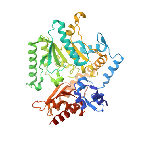 | |
UniProt | |||||
Entity Groups | |||||
| Sequence Clusters | 30% Identity50% Identity70% Identity90% Identity95% Identity100% Identity | ||||
| UniProt Group | P53555 | ||||
Sequence AnnotationsExpand | |||||
Reference Sequence | |||||
| Ligands 2 Unique | |||||
|---|---|---|---|---|---|
| ID | Chains | Name / Formula / InChI Key | 2D Diagram | 3D Interactions | |
| LLP (Subject of Investigation/LOI) Download:Ideal Coordinates CCD File | C [auth A] | (2S)-2-amino-6-[[3-hydroxy-2-methyl-5-(phosphonooxymethyl)pyridin-4-yl]methylideneamino]hexanoic acid C14 H22 N3 O7 P YQSOQJORMNSDJL-QFULYMJESA-N |  | ||
| PMP Download:Ideal Coordinates CCD File | D [auth B] | 4'-DEOXY-4'-AMINOPYRIDOXAL-5'-PHOSPHATE C8 H13 N2 O5 P ZMJGSOSNSPKHNH-UHFFFAOYSA-N |  | ||
| Length ( Å ) | Angle ( ˚ ) |
|---|---|
| a = 58.3 | α = 96.15 |
| b = 60.54 | β = 106.04 |
| c = 62.72 | γ = 99.23 |
| Software Name | Purpose |
|---|---|
| REFMAC | refinement |
| XSCALE | data scaling |
| PDB_EXTRACT | data extraction |
| XDS | data reduction |
| PHASER | phasing |
| Funding Organization | Location | Grant Number |
|---|---|---|
| National Science Foundation (NSF, United States) | United States | 1833181 |


