Optically Modulated and Optically Activated Delayed Fluorescent Proteins through Dark State Engineering
Peng, B., Dikdan, R., Hill, S.E., Patterson-Orazem, A.C., Lieberman, R.L., Fahrni, C.J., Dickson, R.M.(2021) J Phys Chem B 125: 5200-5209
- PubMed: 33978414 Search on PubMedSearch on PubMed Central
- DOI: https://doi.org/10.1021/acs.jpcb.1c00649
- Primary Citation Related Structures:
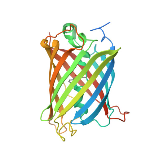
6VIO - PubMed Abstract:
Modulating fluorescent protein emission holds great potential for increasing readout sensitivity for applications in biological imaging and detection. Here, we identify and engineer optically modulated yellow fluorescent proteins (EYFP, originally 10C, but renamed EYFP later, and mVenus) to yield new emitters with distinct modulation profiles and unique, optically gated, delayed fluorescence. The parent YFPs are individually modulatable through secondary illumination, depopulating a long-lived dark state to dynamically increase fluorescence. A single point mutation introduced near the chromophore in each of these YFPs provides access to a second, even longer-lived modulatable dark state, while a different double mutant renders EYFP unmodulatable. The naturally occurring dark state in the parent YFPs yields strong fluorescence modulation upon long-wavelength-induced dark state depopulation, allowing selective detection at the frequency at which the long wavelength secondary laser is intensity modulated. Distinct from photoswitches, however, this near IR secondary coexcitation repumps the emissive S 1 level from the long-lived triplet state, resulting in optically activated delayed fluorescence (OADF). This OADF results from secondary laser-induced, reverse intersystem crossing (RISC), producing additional nanosecond-lived, visible fluorescence that is delayed by many microseconds after the primary excitation has turned off. Mutation of the parent chromophore environment opens an additional modulation pathway that avoids the OADF-producing triplet state, resulting in a second, much longer-lived, modulatable dark state. These Optically Modulated and Optically Activated Delayed Fluorescent Proteins (OMFPs and OADFPs) are thus excellent for background- and reference-free, high sensitivity cellular imaging, but time-gated OADF offers a second modality for true background-free detection. Our combined structural and spectroscopic data not only gives additional mechanistic details for designing optically modulated fluorescent proteins but also provides the opportunity to distinguish similarly emitting OMFPs through OADF and through their unique modulation spectra.
- School of Chemistry & Biochemistry and Petit Institute for Biosciences and Bioengineering, Georgia Institute of Technology, Atlanta, Georgia 30332-0400, United States.
Organizational Affiliation: