6VEQ | pdb_00006veq

Con-Ins G1 in complex with the human insulin microreceptor in turn in complex with Fv 83-7


Experimental Data Snapshot

  • Method: X-RAY DIFFRACTION
  • Resolution: 3.25 Å
  • R-Value Free: 
    0.277 (Depositor), 0.276 (DCC) 
  • R-Value Work: 
    0.228 (Depositor), 0.235 (DCC) 
  • R-Value Observed: 
    0.230 (Depositor) 

Starting Model: experimental
View more details

wwPDB Validation 3D Report Full Report

Validation slider image for 6VEQ

Ligand Structure Quality Assessment 


This is version 2.2 of the entry. See complete history. 

Literature

A structurally minimized yet fully active insulin based on cone-snail venom insulin principles.

Xiong, X., Menting, J.G., Disotuar, M.M., Smith, N.A., Delaine, C.A., Ghabash, G., Agrawal, R., Wang, X., He, X., Fisher, S.J., MacRaild, C.A., Norton, R.S., Gajewiak, J., Forbes, B.E., Smith, B.J., Safavi-Hemami, H., Olivera, B., Lawrence, M.C., Chou, D.H.

(2020) Nat Struct Mol Biol 27: 615-624

  • DOI: https://doi.org/10.1038/s41594-020-0430-8
  • Primary Citation Related Structures: 
    6VEP, 6VEQ, 6VET

  • PubMed Abstract: 

    Human insulin and its current therapeutic analogs all show propensity, albeit varyingly, to self-associate into dimers and hexamers, which delays their onset of action and makes blood glucose management difficult for people with diabetes. Recently, we described a monomeric, insulin-like peptide in cone-snail venom with moderate human insulin-like bioactivity. Here, with insights from structural biology studies, we report the development of mini-Ins-a human des-octapeptide insulin analog-as a structurally minimal, full-potency insulin. Mini-Ins is monomeric and, despite the lack of the canonical B-chain C-terminal octapeptide, has similar receptor binding affinity to human insulin. Four mutations compensate for the lack of contacts normally made by the octapeptide. Mini-Ins also has similar in vitro insulin signaling and in vivo bioactivities to human insulin. The full bioactivity of mini-Ins demonstrates the dispensability of the PheB24-PheB25-TyrB26 aromatic triplet and opens a new direction for therapeutic insulin development.


  • Organizational Affiliation: 
    • Department of Biochemistry, University of Utah, Salt Lake City, UT, USA.

Macromolecule Content 

  • Total Structure Weight: 148.16 kDa 
  • Atom Count: 9,267 
  • Modeled Residue Count: 1,134 
  • Deposited Residue Count: 1,270 
  • Unique protein chains: 6

Macromolecules

Find similar proteins by:  Sequence   |   3D Structure  
Entity ID: 1
MoleculeChains  Sequence LengthOrganismDetailsImage
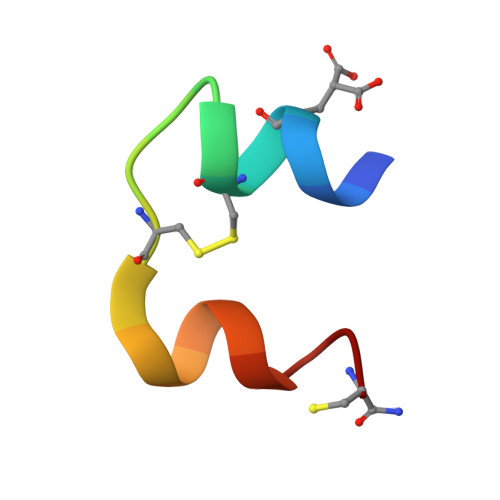
Con-Ins G1a A chainA,
L [auth G]
20Conus geographusMutation(s): 0 
UniProt
Find proteins for A0A0B5AC95 (Conus geographus)
Explore A0A0B5AC95 
Go to UniProtKB:  A0A0B5AC95
Entity Groups
Sequence Clusters30% Identity50% Identity70% Identity90% Identity95% Identity100% Identity
UniProt GroupA0A0B5AC95
Sequence Annotations
Expand
Reference Sequence
Find similar proteins by:  Sequence   |   3D Structure  
Entity ID: 2
MoleculeChains  Sequence LengthOrganismDetailsImage
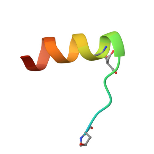
Con-Ins G1 B chainB,
K [auth H]
23Conus geographusMutation(s): 0 
UniProt
Find proteins for A0A0B5A8Q2 (Conus geographus)
Explore A0A0B5A8Q2 
Go to UniProtKB:  A0A0B5A8Q2
Entity Groups
Sequence Clusters30% Identity50% Identity70% Identity90% Identity95% Identity100% Identity
UniProt GroupA0A0B5A8Q2
Sequence Annotations
Expand
Reference Sequence
Find similar proteins by:|  3D Structure
Entity ID: 3
MoleculeChains  Sequence LengthOrganismDetailsImage
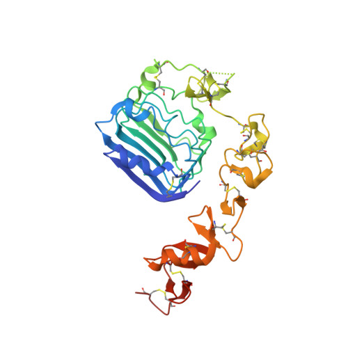
Insulin receptor subunit alphaC [auth E],
G [auth K]
317Homo sapiensMutation(s): 0 
Gene Names: INSR
EC: 2.7.10.1
UniProt & NIH Common Fund Data Resources
Find proteins for P06213 (Homo sapiens)
Explore P06213 
Go to UniProtKB:  P06213
PHAROS:  P06213
GTEx:  ENSG00000171105 
Entity Groups
Sequence Clusters30% Identity50% Identity70% Identity90% Identity95% Identity100% Identity
UniProt GroupP06213
Glycosylation
Glycosylation Sites: 5Go to GlyGen: P06213-1
Sequence Annotations
Expand
Reference Sequence
Find similar proteins by:  Sequence   |   3D Structure  
Entity ID: 4
MoleculeChains  Sequence LengthOrganismDetailsImage
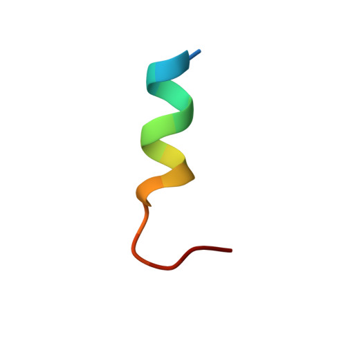
Insulin receptorD [auth F],
H [auth L]
16Homo sapiensMutation(s): 0 
EC: 2.7.10.1
UniProt & NIH Common Fund Data Resources
Find proteins for P06213 (Homo sapiens)
Explore P06213 
Go to UniProtKB:  P06213
PHAROS:  P06213
GTEx:  ENSG00000171105 
Entity Groups
Sequence Clusters30% Identity50% Identity70% Identity90% Identity95% Identity100% Identity
UniProt GroupP06213
Sequence Annotations
Expand
Reference Sequence
Find similar proteins by:|  3D Structure
Entity ID: 5
MoleculeChains  Sequence LengthOrganismDetailsImage
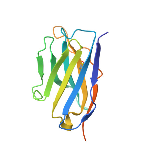
Fv 83-7 heavy chainE [auth C],
I
138Mus musculusMutation(s): 0 
Entity Groups
Sequence Clusters30% Identity50% Identity70% Identity90% Identity95% Identity100% Identity
Sequence Annotations
Expand
Reference Sequence
Find similar proteins by:|  3D Structure
Entity ID: 6
MoleculeChains  Sequence LengthOrganismDetailsImage
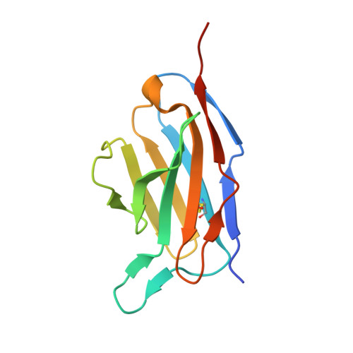
Fv 83-7 light chainF [auth D],
J
121Mus musculusMutation(s): 0 
Entity Groups
Sequence Clusters30% Identity50% Identity70% Identity90% Identity95% Identity100% Identity
Sequence Annotations
Expand
Reference Sequence

Oligosaccharides

Help  
Entity ID: 7
MoleculeChains Length2D Diagram GlycosylationD Interactions
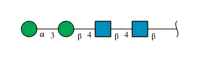
alpha-D-mannopyranose-(1-3)-beta-D-mannopyranose-(1-4)-2-acetamido-2-deoxy-beta-D-glucopyranose-(1-4)-2-acetamido-2-deoxy-beta-D-glucopyranose
M, O
4N-Glycosylation
Glycosylation Resources
GlyTouCan: G81315DD
GlyCosmos: G81315DD
GlyGen: G81315DD
Entity ID: 8
MoleculeChains Length2D Diagram GlycosylationD Interactions
beta-D-mannopyranose-(1-4)-2-acetamido-2-deoxy-beta-D-glucopyranose-(1-4)-[alpha-L-fucopyranose-(1-6)]2-acetamido-2-deoxy-beta-D-glucopyranose
N, P
4N-Glycosylation
Glycosylation Resources
GlyTouCan: G32152BH
GlyCosmos: G32152BH
GlyGen: G32152BH

Small Molecules

Ligands 2 Unique
IDChains Name / Formula / InChI Key2D Diagram3D Interactions
NAG

Query on NAG


Download:Ideal Coordinates CCD File
Q [auth E]
R [auth E]
S [auth E]
W [auth K]
X [auth K]
Q [auth E],
R [auth E],
S [auth E],
W [auth K],
X [auth K],
Y [auth K]
2-acetamido-2-deoxy-beta-D-glucopyranose
C8 H15 N O6
OVRNDRQMDRJTHS-FMDGEEDCSA-N
SO4

Query on SO4


Download:Ideal Coordinates CCD File
T [auth E],
U [auth E],
V [auth D],
Z [auth K]
SULFATE ION
O4 S
QAOWNCQODCNURD-UHFFFAOYSA-L
Modified Residues  3 Unique
IDChains TypeFormula2D DiagramParent
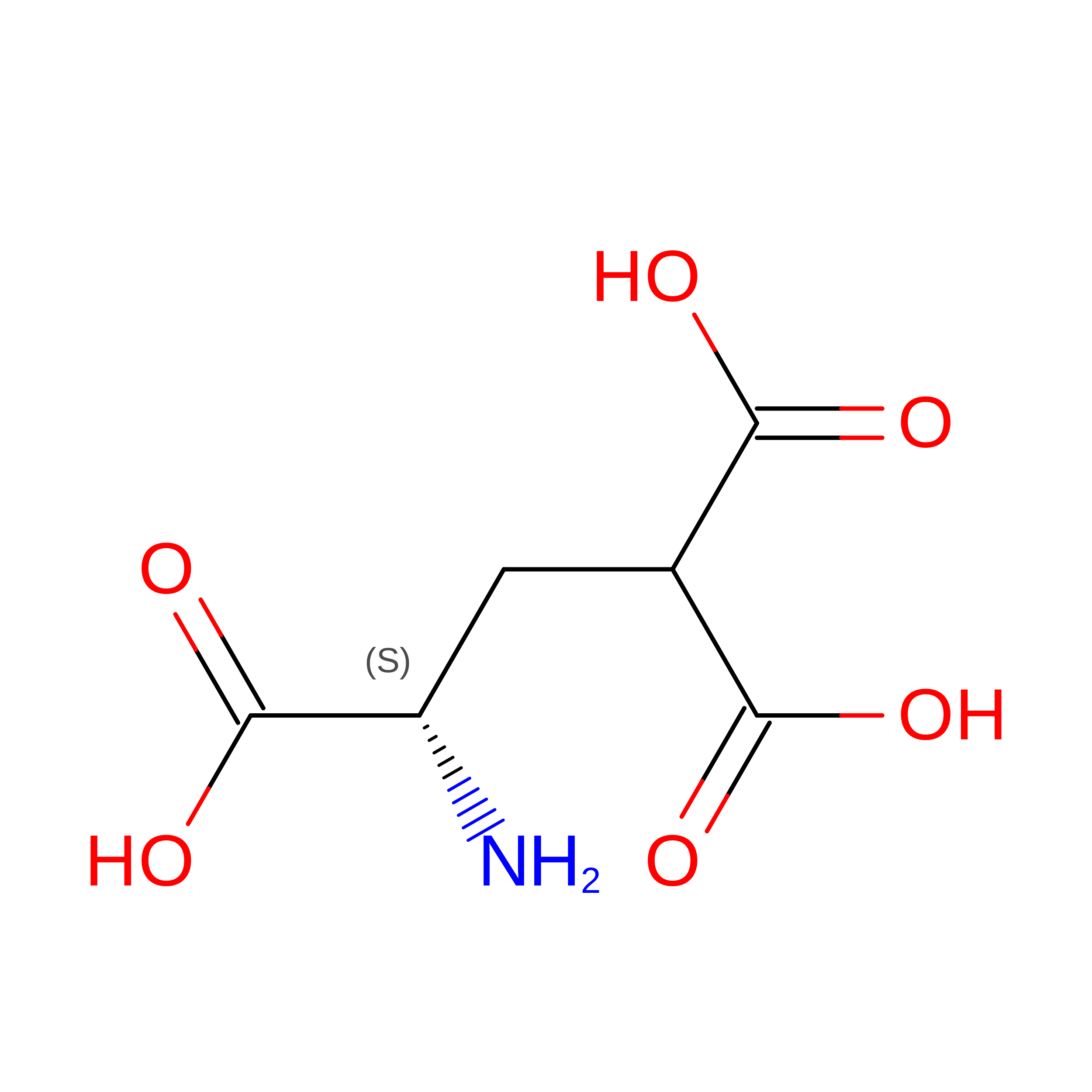
CGU
Query on CGU
A,
L [auth G]
L-PEPTIDE LINKINGC6 H9 N O6GLU
CY3
Query on CY3
A,
L [auth G]
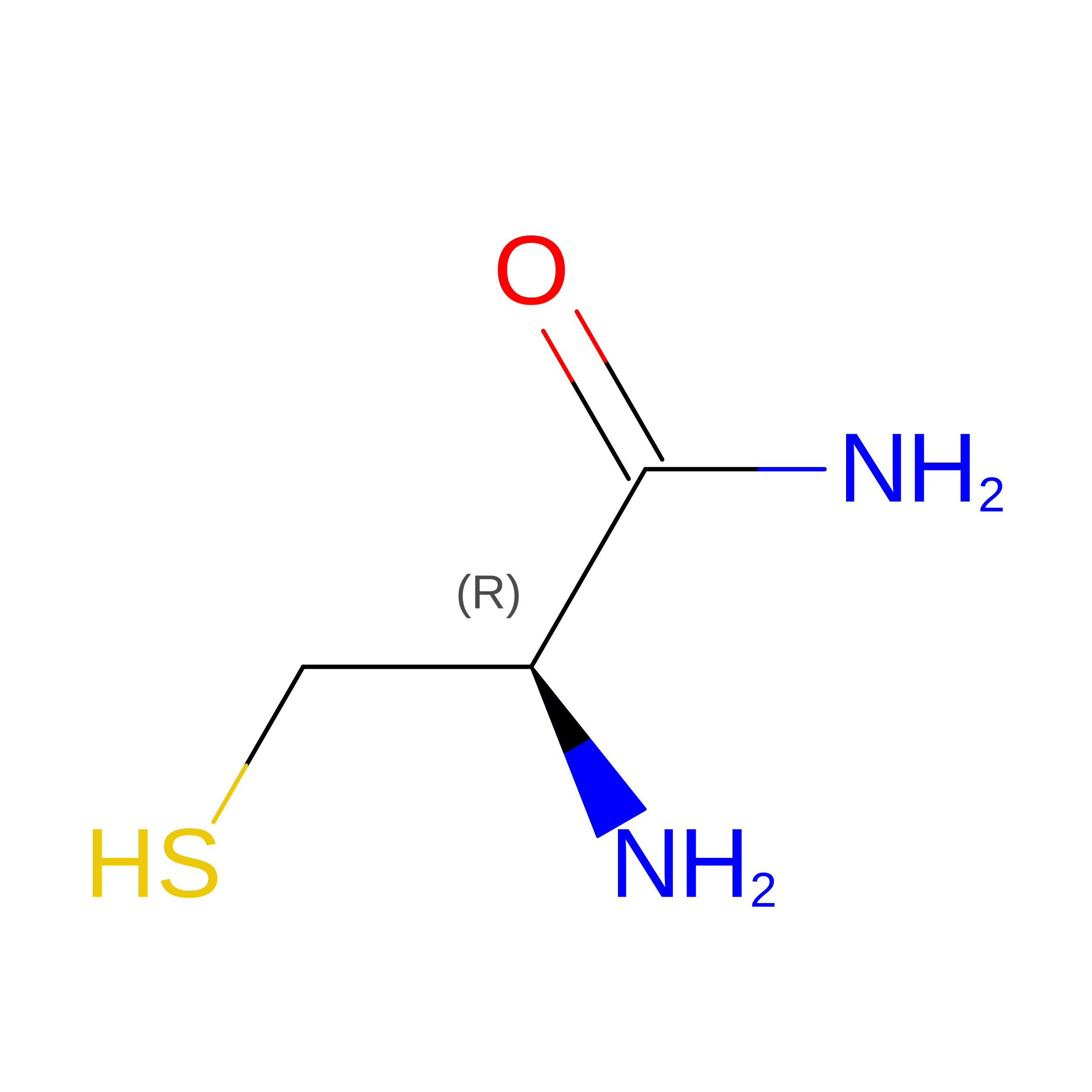
L-PEPTIDE LINKINGC3 H8 N2 O SCYS
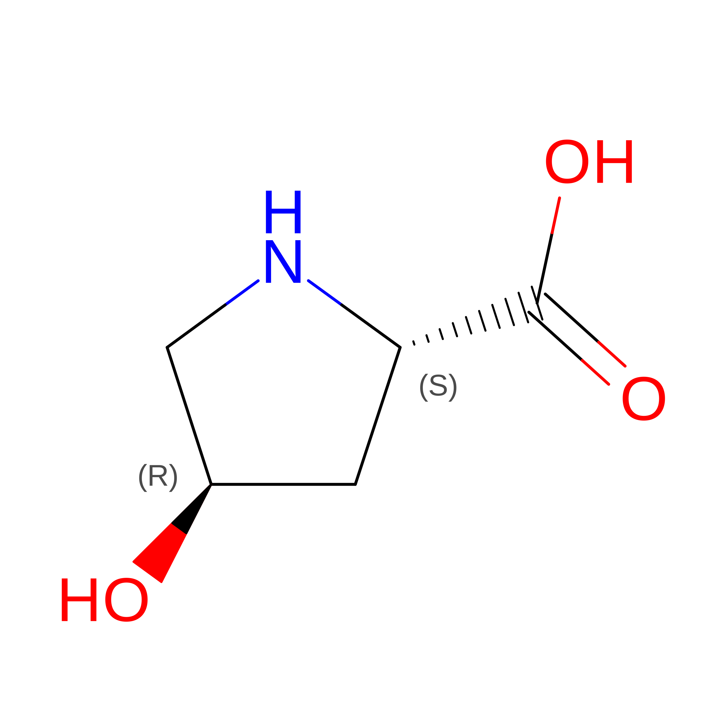
HYP
Query on HYP
B,
K [auth H]
L-PEPTIDE LINKINGC5 H9 N O3PRO

Experimental Data & Validation

Experimental Data

  • Method: X-RAY DIFFRACTION
  • Resolution: 3.25 Å
  • R-Value Free:  0.277 (Depositor), 0.276 (DCC) 
  • R-Value Work:  0.228 (Depositor), 0.235 (DCC) 
  • R-Value Observed: 0.230 (Depositor) 
Space Group: I 2 2 2
Unit Cell:
Length ( Å )Angle ( ˚ )
a = 106.16α = 90
b = 227.12β = 90
c = 228.7γ = 90
Software Package:
Software NamePurpose
PHENIXrefinement
XDSdata reduction
XSCALEdata scaling
PHASERphasing

Structure Validation

View Full Validation Report


Ligand Structure Quality Assessment 


Entry History 

& Funding Information

Deposition Data


Funding OrganizationLocationGrant Number
National Health and Medical Research Council (NHMRC, Australia)AustraliaAPP1143546
Other privateUnited States5-CDA-2018-572-A-N

Revision History  (Full details and data files)

  • Version 1.0: 2020-06-03
    Type: Initial release
  • Version 1.1: 2020-06-17
    Changes: Database references
  • Version 1.2: 2020-07-22
    Changes: Database references
  • Version 2.0: 2020-07-29
    Type: Remediation
    Reason: Carbohydrate remediation
    Changes: Atomic model, Data collection, Derived calculations, Structure summary
  • Version 2.1: 2023-10-11
    Changes: Data collection, Database references, Refinement description, Structure summary
  • Version 2.2: 2023-11-15
    Changes: Data collection