6US5

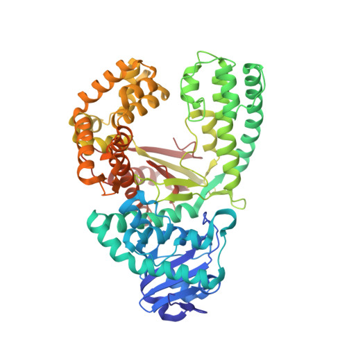
DNA polymerase I Large Fragment from Bacillus stearothermophilus with DNA template, 3'-amino primer, dGpNHpp analog, and Mn2+


Experimental Data Snapshot

  • Method: X-RAY DIFFRACTION
  • Resolution: 2.25 Å
  • R-Value Free: 0.278 
  • R-Value Work: 0.224 
  • R-Value Observed: 0.227 

wwPDB Validation   3D Report Full Report


Ligand Structure Quality Assessment 


This is version 1.2 of the entry. See complete history


Literature

Synthesis of phosphoramidate-linked DNA by a modified DNA polymerase.

Lelyveld, V.S.Zhang, W.Szostak, J.W.

(2020) Proc Natl Acad Sci U S A 117: 7276-7283

  • DOI: https://doi.org/10.1073/pnas.1922400117
  • Primary Citation of Related Structures:  
    6UR2, 6UR4, 6UR9, 6US5

  • PubMed Abstract: 

    All known polymerases copy genetic material by catalyzing phosphodiester bond formation. This highly conserved activity proceeds by a common mechanism, such that incorporated nucleoside analogs terminate chain elongation if the resulting primer strand lacks a terminal hydroxyl group. Even conservatively substituted 3'-amino nucleotides generally act as chain terminators, and no enzymatic pathway for their polymerization has yet been found. Although 3'-amino nucleotides can be chemically coupled to yield stable oligonucleotides containing N3'→P5' phosphoramidate (NP) bonds, no such internucleotide linkages are known to occur in nature. Here, we report that 3'-amino terminated primers are, in fact, slowly extended by the DNA polymerase from B. stearothermophilus in a template-directed manner. When its cofactor is Ca 2+ rather than Mg 2+ , the reaction is fivefold faster, permitting multiple turnover NP bond formation to yield NP-DNA strands from the corresponding 3'-amino-2',3'-dideoxynucleoside 5'-triphosphates. A single active site mutation further enhances the rate of NP-DNA synthesis by an additional 21-fold. We show that DNA-dependent NP-DNA polymerase activity depends on conserved active site residues and propose a likely mechanism for this activity based on a series of crystal structures of bound complexes. Our results significantly broaden the catalytic scope of polymerase activity and suggest the feasibility of a genetic transition between native nucleic acids and NP-DNA.


  • Organizational Affiliation

    Department of Molecular Biology, Center for Computational and Integrative Biology, Massachusetts General Hospital, Boston, MA 02114.


Macromolecules

Find similar proteins by:  (by identity cutoff)  |  3D Structure
Entity ID: 1
MoleculeChains Sequence LengthOrganismDetailsImage
DNA polymerase I
A, D
579Geobacillus stearothermophilusMutation(s): 2 
Gene Names: DPO1polA
EC: 2.7.7.7
UniProt
Find proteins for D9N168 (Geobacillus stearothermophilus)
Explore D9N168 
Go to UniProtKB:  D9N168
Entity Groups  
Sequence Clusters30% Identity50% Identity70% Identity90% Identity95% Identity100% Identity
UniProt GroupD9N168
Sequence Annotations
Expand
  • Reference Sequence

Find similar nucleic acids by:  Sequence   |   3D Structure  

Entity ID: 2
MoleculeChains LengthOrganismImage
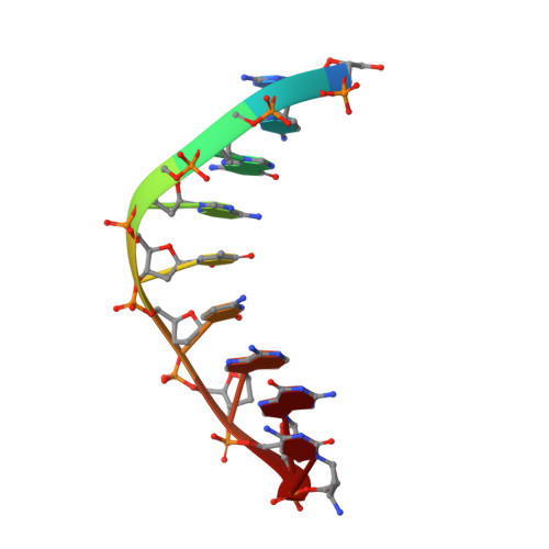
DNA (5'-D(*GP*CP*GP*AP*TP*CP*AP*GP*(C42))-3')
B, E
9synthetic construct
Sequence Annotations
Expand
  • Reference Sequence

Find similar nucleic acids by:  Sequence   |   3D Structure  

Entity ID: 3
MoleculeChains LengthOrganismImage
DNA (5'-D(*CP*AP*CP*GP*CP*TP*GP*AP*TP*CP*GP*CP*A)-3')
C, F
13synthetic construct
Sequence Annotations
Expand
  • Reference Sequence
Small Molecules
Ligands 3 Unique
IDChains Name / Formula / InChI Key2D Diagram3D Interactions
XG4
Query on XG4

Download Ideal Coordinates CCD File 
G [auth A],
N [auth D]
2'-deoxy-5'-O-[(R)-hydroxy{[(R)-hydroxy(phosphonooxy)phosphoryl]amino}phosphoryl]guanosine
C10 H17 N6 O12 P3
DWGAAFQEGIMTIA-KVQBGUIXSA-N
SO4
Query on SO4

Download Ideal Coordinates CCD File 
I [auth A]
J [auth A]
K [auth A]
L [auth A]
M [auth C]
I [auth A],
J [auth A],
K [auth A],
L [auth A],
M [auth C],
P [auth D],
Q [auth D]
SULFATE ION
O4 S
QAOWNCQODCNURD-UHFFFAOYSA-L
MN
Query on MN

Download Ideal Coordinates CCD File 
H [auth A],
O [auth D]
MANGANESE (II) ION
Mn
WAEMQWOKJMHJLA-UHFFFAOYSA-N
Experimental Data & Validation

Experimental Data

  • Method: X-RAY DIFFRACTION
  • Resolution: 2.25 Å
  • R-Value Free: 0.278 
  • R-Value Work: 0.224 
  • R-Value Observed: 0.227 
  • Space Group: P 21 21 21
Unit Cell:
Length ( Å )Angle ( ˚ )
a = 93.761α = 90
b = 108.805β = 90
c = 149.969γ = 90
Software Package:
Software NamePurpose
PHENIXrefinement
PDB_EXTRACTdata extraction
HKL-2000data reduction
HKL-2000data scaling
PHASERphasing

Structure Validation

View Full Validation Report


Ligand Structure Quality Assessment 


Entry History & Funding Information

Deposition Data


Funding OrganizationLocationGrant Number
Howard Hughes Medical Institute (HHMI)United States--

Revision History  (Full details and data files)

  • Version 1.0: 2020-03-18
    Type: Initial release
  • Version 1.1: 2020-05-27
    Changes: Database references
  • Version 1.2: 2023-10-11
    Changes: Data collection, Database references, Derived calculations, Refinement description