Primary Citation Related Structures: 6SNC, 6SND, 6SNE
PubMed Abstract:
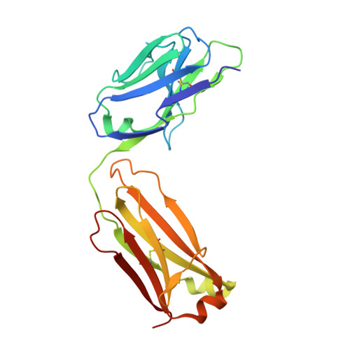
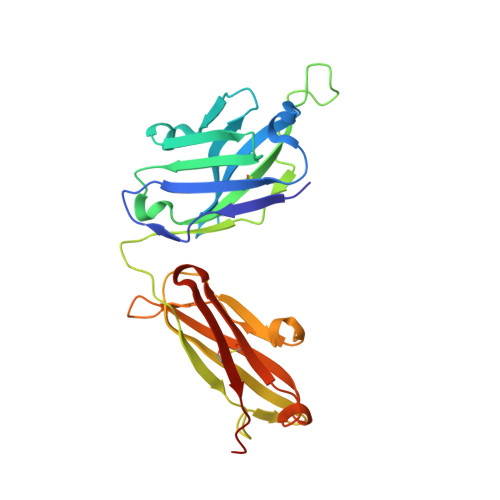
Potent and broadly neutralizing antibodies (bnAbs) are the hallmark of HIV-1 protection by vaccination. The membrane-proximal external region (MPER) of the HIV-1 gp41 fusion protein is targeted by the most broadly reactive HIV-1 neutralizing antibodies. Here, we examine the structural and molecular mechansims of neutralization by anti-MPER bnAb, LN01, which was isolated from lymph-node-derived germinal center B cells of an elite controller and exhibits broad neutralization breadth. LN01 engages both MPER and the transmembrane (TM) region, which together form a continuous helix in complex with LN01. The tilted TM orientation allows LN01 to interact simultaneously with the peptidic component of the MPER epitope and membrane via two specific lipid binding sites of the antibody paratope. Although LN01 carries a high load of somatic mutations, most key residues interacting with the MPER epitope and lipids are germline encoded, lending support for the LN01 epitope as a candidate for lineage-based vaccine development.
Organizational Affiliation:
Institute for Research in Biomedicine, Bellinzona 6500, Ticino, Switzerland.
Swiss Vaccine Research Institute, Lausanne University Hospital, University of Lausanne, 1011 Lausanne, Switzerland.
Institut de Biologie Structurale (IBS), University Grenoble Alpes, CEA, CNRS, 38000 Grenoble, France.
LPCT, UMR 7019 Université de Lorraine CNRS, 54500 Vandœuvre-lès-Nancy, France; Laboratoire International Associé CNRS and University of Illinois at Urbana-Champaign, LPCT, UMR 7019 Universiteé de Lorraine CNRS, Vandœuvre-lès-Nancy 54500, France.
LPCT, UMR 7019 Université de Lorraine CNRS, 54500 Vandœuvre-lès-Nancy, France; Laboratoire International Associé CNRS and University of Illinois at Urbana-Champaign, LPCT, UMR 7019 Universiteé de Lorraine CNRS, Vandœuvre-lès-Nancy 54500, France; Department of Physics, University of Illinois at Urbana-Champaign, Urbana, IL 61801, USA.
Humabs Biomed SA, Vir Biotechnology, 6500 Bellinzona, Ticino, Switzerland.
Duke Human Vaccine Institute, Durham, NC 27710, USA; Paris Diderot University, Sorbonne Paris Cité, Paris 75013, France.
Duke Human Vaccine Institute, Durham, NC 27710, USA.
Department of Biomedical and Clinical Sciences, Luigi Sacco University Hospital, Università di Milano, 20157 Milan, Italy; III Division of Infectious Diseases, ASST Fatebenefratelli-Sacco, 20157 Milan, Italy.
Department of Biomedical and Clinical Sciences, Luigi Sacco University Hospital, Università di Milano, 20157 Milan, Italy.
Institut Pasteur, Virus & Immunity Unit, CNRS UMR 3569, Paris 75015, France; Vaccine Research Institute, 94000 Créteil, France.
Institut Pasteur, Virus & Immunity Unit, CNRS UMR 3569, Paris 75015, France; Vaccine Research Institute, 94000 Créteil, France; Paris Diderot University, Sorbonne Paris Cité, Paris 75013, France.
Beth Israel Deaconess Medical Center, Harvard Medical School, Boston, MA 02215, USA.
Department of Surgery, Duke University Medical Center, Durham, NC 27710, USA.
Humabs Biomed SA, Vir Biotechnology, 6500 Bellinzona, Ticino, Switzerland. Electronic address: dcorti@vir.bio.
Swiss Vaccine Research Institute, Lausanne University Hospital, University of Lausanne, 1011 Lausanne, Switzerland; Service of Immunology and Allergy, Lausanne University Hospital, University of Lausanne, 1011 Lausanne, Switzerland. Electronic address: giuseppe.pantaleo@chuv.ch.
Institut de Biologie Structurale (IBS), University Grenoble Alpes, CEA, CNRS, 38000 Grenoble, France. Electronic address: winfried.weissenhorn@ibs.fr.