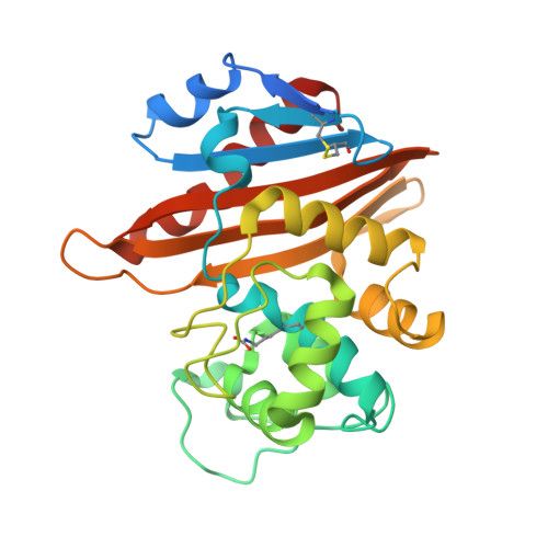
Structural insights into the enhanced carbapenemase efficiency of OXA-655 compared to OXA-10.
Leiros, H.S., Thomassen, A.M., Samuelsen, O., Flach, C.F., Kotsakis, S.D., Larsson, D.G.J.(2020) FEBS Open Bio 10: 1821-1832
- PubMed: 32683794 Search on PubMedSearch on PubMed Central
- DOI: https://doi.org/10.1002/2211-5463.12935
- Primary Citation Related Structures:
6SKP, 6SKQ, 6SKR - PubMed Abstract:
Carbapenemases are the main cause of carbapenem resistance in Gram-negative bacteria. How β-lactamases with weak carbapenemase activity, such as the OXA-10-type class D β-lactamases, contribute to anti-bacterial drug resistance is unclear. OXA-655 is a T26M and V117L OXA-10 variant, recently identified from hospital wastewater. Despite exhibiting stronger carbapenemase activity towards ertapenem (ETP) and meropenem (MEM) in Escherichia coli, OXA-655 exhibits reduced activity towards oxyimino-substituted β-lactams like ceftazidime. Here, we have solved crystal structures of OXA-10 in complex with imipenem (IPM) and ETP, and OXA-655 in complex with MEM in order to unravel the structure-function relationship and the impact of residue 117 in enzyme catalysis. The new crystal structures show that L117 is situated at a critical position with enhanced Van der Waals interactions to L155 in the omega loop. This restricts the movements of L155 and could explain the reduced ability for OXA-655 to bind a bulky oxyimino group. The V117L replacement in OXA-655 makes the active site S67 and the carboxylated K70 more water exposed. This could affect the supply of new deacylation water molecules required for hydrolysis and possibly the carboxylation rate of K70. But most importantly, L117 leaves more space for binding of the hydroxyethyl group in carbapenems. In summary, the crystal structures highlight the importance of residue 117 in OXA-10 variants for carbapenemase activity. This study also illustrates the impact of a single amino acid substitution on the substrate profile of OXA-10 and the evolutionary potential of new OXA-10 variants.
- The Norwegian Structural Biology Centre (NorStruct), Department of Chemistry, Faculty of Science and Technology, UiT The Arctic University of Norway, Tromsø, Norway.
Organizational Affiliation: