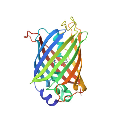
6QUJ

GHK tagged GFP variant


Experimental Data Snapshot

  • Method: X-RAY DIFFRACTION
  • Resolution: 1.68 Å
  • R-Value Free: 0.221 
  • R-Value Work: 0.172 
  • R-Value Observed: 0.174 

wwPDB Validation   3D Report Full Report


This is version 3.0 of the entry. See complete history


Literature

The copper(II)-binding tripeptide GHK, a valuable crystallization and phasing tag for macromolecular crystallography.

Mehr, A.Henneberg, F.Chari, A.Gorlich, D.Huyton, T.

(2020) Acta Crystallogr D Struct Biol 76: 1222-1232

  • DOI: https://doi.org/10.1107/S2059798320013741
  • Primary Citation of Related Structures:  
    6QUG, 6QUH, 6QUI, 6QUJ

  • PubMed Abstract: 

    The growth of diffraction-quality crystals and experimental phasing remain two of the main bottlenecks in protein crystallography. Here, the high-affinity copper(II)-binding tripeptide GHK was fused to the N-terminus of a GFP variant and an MBP-FG peptide fusion. The GHK tag promoted crystallization, with various residues (His, Asp, His/Pro) from symmetry molecules completing the copper(II) square-pyramidal coordination sphere. Rapid structure determination by copper SAD phasing could be achieved, even at a very low Bijvoet ratio or after significant radiation damage. When collecting highly redundant data at a wavelength close to the copper absorption edge, residual S-atom positions could also be located in log-likelihood-gradient maps and used to improve the phases. The GHK copper SAD method provides a convenient way of both crystallizing and phasing macromolecular structures, and will complement the current trend towards native sulfur SAD and MR-SAD phasing.


  • Organizational Affiliation

    Department of Structural Dynamics, Max Planck Institute for Biophysical Chemistry, Göttingen, Germany.


Macromolecules
Find similar proteins by:  (by identity cutoff)  |  3D Structure
Entity ID: 1
MoleculeChains Sequence LengthOrganismDetailsImage
Green fluorescent proteinA [auth B],
B [auth E]
229Aequorea victoriaMutation(s): 0 
Gene Names: GFP
UniProt
Find proteins for P42212 (Aequorea victoria)
Explore P42212 
Go to UniProtKB:  P42212
Entity Groups  
Sequence Clusters30% Identity50% Identity70% Identity90% Identity95% Identity100% Identity
UniProt GroupP42212
Sequence Annotations
Expand
  • Reference Sequence
Small Molecules
Modified Residues  1 Unique
IDChains TypeFormula2D DiagramParent
CRO
Query on CRO
A [auth B],
B [auth E]
L-PEPTIDE LINKINGC15 H17 N3 O5THR, TYR, GLY
Experimental Data & Validation

Experimental Data

  • Method: X-RAY DIFFRACTION
  • Resolution: 1.68 Å
  • R-Value Free: 0.221 
  • R-Value Work: 0.172 
  • R-Value Observed: 0.174 
  • Space Group: P 65 2 2
Unit Cell:
Length ( Å )Angle ( ˚ )
a = 85.99α = 90
b = 85.99β = 90
c = 270.14γ = 120
Software Package:
Software NamePurpose
REFMACrefinement
XDSdata reduction
Aimlessdata scaling
CRANK2phasing

Structure Validation

View Full Validation Report


Entry History 

Deposition Data

Revision History  (Full details and data files)

  • Version 1.0: 2020-05-27
    Type: Initial release
  • Version 2.0: 2020-12-09
    Type: Coordinate replacement
    Reason: Atomic clashes
    Changes: Advisory, Atomic model, Author supporting evidence, Data collection, Database references, Derived calculations, Non-polymer description, Refinement description, Structure summary
  • Version 2.1: 2020-12-16
    Changes: Database references
  • Version 3.0: 2022-12-07
    Changes: Advisory, Atomic model, Data collection, Database references, Derived calculations, Polymer sequence, Refinement description, Source and taxonomy, Structure summary