6PRV

58nt RNA L11-binding domain from E. coli 23S rRNA


Experimental Data Snapshot

  • Method: X-RAY DIFFRACTION
  • Resolution: 2.71 Å
  • R-Value Free: 0.260 
  • R-Value Work: 0.204 
  • R-Value Observed: 0.207 

wwPDB Validation   3D Report Full Report


This is version 1.2 of the entry. See complete history


Literature

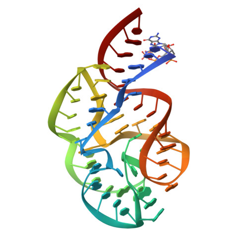
Ribosomal Protein L11 Selectively Stabilizes a Tertiary Structure of the GTPase Center rRNA Domain.

Welty, R.Rau, M.Pabit, S.Dunstan, M.S.Conn, G.L.Pollack, L.Hall, K.B.

(2020) J Mol Biol 432: 991-1007

  • DOI: https://doi.org/10.1016/j.jmb.2019.12.010
  • Primary Citation of Related Structures:  
    6PRV

  • PubMed Abstract: 

    The GTPase Center (GAC) RNA domain in bacterial 23S rRNA is directly bound by ribosomal protein L11, and this complex is essential to ribosome function. Previous cocrystal structures of the 58-nucleotide GAC RNA bound to L11 revealed the intricate tertiary fold of the RNA domain, with one monovalent and several divalent ions located in specific sites within the structure. Here, we report a new crystal structure of the free GAC that is essentially identical to the L11-bound structure, which retains many common sites of divalent ion occupation. This new structure demonstrates that RNA alone folds into its tertiary structure with bound divalent ions. In solution, we find that this tertiary structure is not static, but rather is best described as an ensemble of states. While L11 protein cannot bind to the GAC until the RNA has adopted its tertiary structure, new experimental data show that L11 binds to Mg 2+ -dependent folded states, which we suggest lie along the folding pathway of the RNA. We propose that L11 stabilizes a specific GAC RNA tertiary state, corresponding to the crystal structure, and that this structure reflects the functionally critical conformation of the rRNA domain in the fully assembled ribosome.


  • Organizational Affiliation

    Department of Biochemistry and Molecular Biophysics, Washington University School of Medicine, 660 S Euclid Ave, St Louis, MO, 63110, USA; Department of Chemistry, University of Michigan, Ann Arbor, MI, 48109, USA.


Macromolecules
Find similar nucleic acids by:  (by identity cutoff)  |  3D Structure
Entity ID: 1
MoleculeChains LengthOrganismImage
23S rRNA
A, B, C, D
58Escherichia coli
Sequence Annotations
Expand
  • Reference Sequence
Small Molecules
Ligands 2 Unique
IDChains Name / Formula / InChI Key2D Diagram3D Interactions
K (Subject of Investigation/LOI)
Query on K

Download Ideal Coordinates CCD File 
AA [auth C],
N [auth A],
T [auth B]
POTASSIUM ION
K
NPYPAHLBTDXSSS-UHFFFAOYSA-N
MG (Subject of Investigation/LOI)
Query on MG

Download Ideal Coordinates CCD File 
BA [auth D]
CA [auth D]
E [auth A]
F [auth A]
G [auth A]
BA [auth D],
CA [auth D],
E [auth A],
F [auth A],
G [auth A],
H [auth A],
I [auth A],
J [auth A],
K [auth A],
L [auth A],
M [auth A],
O [auth B],
P [auth B],
Q [auth B],
R [auth B],
S [auth B],
U [auth C],
V [auth C],
W [auth C],
X [auth C],
Y [auth C],
Z [auth C]
MAGNESIUM ION
Mg
JLVVSXFLKOJNIY-UHFFFAOYSA-N
Experimental Data & Validation

Experimental Data

  • Method: X-RAY DIFFRACTION
  • Resolution: 2.71 Å
  • R-Value Free: 0.260 
  • R-Value Work: 0.204 
  • R-Value Observed: 0.207 
  • Space Group: C 1 2 1
Unit Cell:
Length ( Å )Angle ( ˚ )
a = 89.61α = 90
b = 73.61β = 100.961
c = 130.28γ = 90
Software Package:
Software NamePurpose
XDSdata reduction
XSCALEdata scaling
PHENIXrefinement
PHENIXphasing

Structure Validation

View Full Validation Report


Entry History 

Deposition Data

Revision History  (Full details and data files)

  • Version 1.0: 2020-01-08
    Type: Initial release
  • Version 1.1: 2020-03-11
    Changes: Database references
  • Version 1.2: 2023-10-11
    Changes: Data collection, Database references, Derived calculations, Refinement description