6MDR | pdb_00006mdr

Cryo-EM structure of the Ceru+32/GFP-17 protomer


Experimental Data Snapshot

  • Method: ELECTRON MICROSCOPY
  • Resolution: 3.47 Å
  • Aggregation State: PARTICLE 
  • Reconstruction Method: SINGLE PARTICLE 

Starting Model: experimental
View more details

wwPDB Validation 3D Report Full Report

Validation slider image for 6MDR

This is version 1.4 of the entry. See complete history

Literature

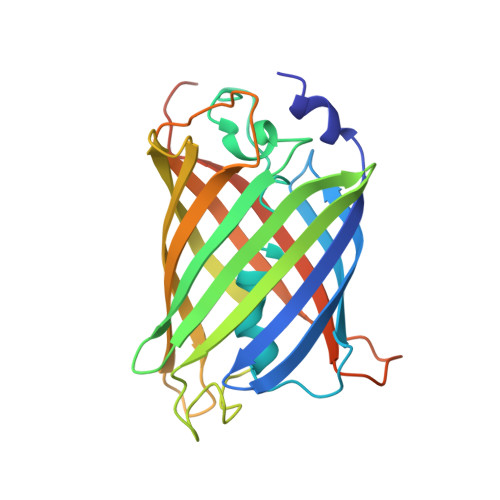
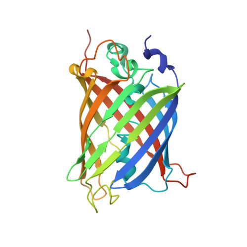
Supercharging enables organized assembly of synthetic biomolecules.

Simon, A.J.Zhou, Y.Ramasubramani, V.Glaser, J.Pothukuchy, A.Gollihar, J.Gerberich, J.C.Leggere, J.C.Morrow, B.R.Jung, C.Glotzer, S.C.Taylor, D.W.Ellington, A.D.

(2019) Nat Chem 11: 204-212

  • DOI: https://doi.org/10.1038/s41557-018-0196-3
  • Primary Citation Related Structures: 
    6MDR

  • PubMed Abstract: 

    Symmetrical protein oligomers are ubiquitous in biological systems and perform key structural and regulatory functions. However, there are few methods for constructing such oligomers. Here we have engineered completely synthetic, symmetrical oligomers by combining pairs of oppositely supercharged variants of a normally monomeric model protein through a strategy we term 'supercharged protein assembly' (SuPrA). We show that supercharged variants of green fluorescent protein can assemble into a variety of architectures including a well-defined symmetrical 16-mer structure that we solved using cryo-electron microscopy at 3.47 Å resolution. The 16-mer is composed of two stacked rings of octamers, in which the octamers contain supercharged proteins of alternating charges, and interactions within and between the rings are mediated by a variety of specific electrostatic contacts. The ready assembly of this structure suggests that combining oppositely supercharged pairs of protein variants may provide broad opportunities for generating novel architectures via otherwise unprogrammed interactions.


  • Organizational Affiliation
    • Center for Systems and Synthetic Biology, University of Texas at Austin, Austin, TX, USA.

Macromolecule Content 

  • Total Structure Weight: 445.21 kDa 
  • Atom Count: 29,456 
  • Modeled Residue Count: 3,696 
  • Deposited Residue Count: 3,896 
  • Unique protein chains: 2

Macromolecules

Find similar proteins by:|  3D Structure
Entity ID: 1
MoleculeChains  Sequence LengthOrganismDetailsImage
Ceru+32247Aequorea victoriaMutation(s): 39 
Gene Names: GFP
UniProt
Find proteins for P42212 (Aequorea victoria)
Explore P42212 
Go to UniProtKB:  P42212
Entity Groups
Sequence Clusters30% Identity50% Identity70% Identity90% Identity95% Identity100% Identity
UniProt GroupP42212
Sequence Annotations
Expand
Reference Sequence
Find similar proteins by:|  3D Structure
Entity ID: 2
MoleculeChains  Sequence LengthOrganismDetailsImage
GFP-17240Aequorea victoriaMutation(s): 22 
Gene Names: GFP
UniProt
Find proteins for P42212 (Aequorea victoria)
Explore P42212 
Go to UniProtKB:  P42212
Entity Groups
Sequence Clusters30% Identity50% Identity70% Identity90% Identity95% Identity100% Identity
UniProt GroupP42212
Sequence Annotations
Expand
Reference Sequence

Experimental Data & Validation

Experimental Data

  • Method: ELECTRON MICROSCOPY
  • Resolution: 3.47 Å
  • Aggregation State: PARTICLE 
  • Reconstruction Method: SINGLE PARTICLE 
EM Software:
TaskSoftware PackageVersion
MODEL REFINEMENTPHENIX
RECONSTRUCTIONRELION

Structure Validation

View Full Validation Report


Entry History 

& Funding Information

Deposition Data


Funding OrganizationLocationGrant Number
Other governmentUnited StatesW911NF-1-51-0120

Revision History  (Full details and data files)

  • Version 1.0: 2019-01-23
    Type: Initial release
  • Version 1.1: 2019-01-30
    Changes: Data collection, Database references
  • Version 1.2: 2019-03-06
    Changes: Data collection, Database references
  • Version 1.3: 2019-04-17
    Changes: Author supporting evidence, Data collection
  • Version 1.4: 2024-03-13
    Changes: Data collection, Database references, Refinement description