6M5Y

Structure of human galectin-1 tandem-repeat mutant with lactose


Experimental Data Snapshot

  • Method: X-RAY DIFFRACTION
  • Resolution: 1.38 Å
  • R-Value Free: 0.219 
  • R-Value Work: 0.183 
  • R-Value Observed: 0.185 

wwPDB Validation   3D Report Full Report


This is version 1.1 of the entry. See complete history


Literature

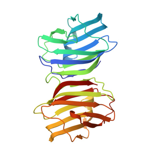
Crystal structure and conformational stability of a galectin-1 tandem-repeat mutant with a short linker.

Nonaka, Y.Ogawa, T.Shoji, H.Nishi, N.Kamitori, S.Nakamura, T.

(2021) Glycobiology 

  • DOI: https://doi.org/10.1093/glycob/cwab101
  • Primary Citation of Related Structures:  
    6M5Y

  • PubMed Abstract: 
  • Modification of the domain architecture of galectins has been attempted to analyze their biological functions and to develop medical applications. Several types of galectin-1 repeat mutants were previously reported but, however, it was not clear whether the native structure of the wild type was retained ...

    Modification of the domain architecture of galectins has been attempted to analyze their biological functions and to develop medical applications. Several types of galectin-1 repeat mutants were previously reported but, however, it was not clear whether the native structure of the wild type was retained. In this study, we determined the crystal structure of a galectin-1 tandem-repeat mutant with a short linker peptide, and compared the unfolding profiles of the wild type and mutant by chemical denaturation. The structure of the mutant was consistent with that of the dimer of the wild type, and both carbohydrate-binding sites were retained. The unfolding curve of the wild type with lactose suggested that the dimer dissociation and the tertiary structure unfolding was concomitant at micromolar protein concentrations. The midpoint denaturant concentration of the wild type was dependent on the protein concentration and lower than that of the mutant. Linking the two subunits significantly stabilized the tertiary structure. The mutant exhibited higher T-cell growth-inhibition activity and comparable hemagglutinating activity. Structural stabilization may prevent the oxidation of the internal cysteine residue.


    Organizational Affiliation

    Department of Endocrinology, Faculty of Medicine, Kagawa University, 1750-1, Ikenobe, Miki-cho, Kita-gun, Kagawa 761-0793, Japan.


Macromolecules
Find similar proteins by:  (by identity cutoff)  |  3D Structure
Entity ID: 1
MoleculeChainsSequence LengthOrganismDetailsImage
Galectin-1,Galectin-1A272Homo sapiensMutation(s): 10 
Gene Names: LGALS1
UniProt & NIH Common Fund Data Resources
Find proteins for P09382 (Homo sapiens)
Explore P09382 
Go to UniProtKB:  P09382
PHAROS:  P09382
Entity Groups  
Sequence Clusters30% Identity50% Identity70% Identity90% Identity95% Identity100% Identity
UniProt GroupP09382
Protein Feature View
Expand
  • Reference Sequence
Oligosaccharides

Help

Entity ID: 2
MoleculeChainsChain Length2D DiagramGlycosylation3D Interactions
beta-D-galactopyranose-(1-4)-beta-D-glucopyranoseB 2N/A Oligosaccharides Interaction
Glycosylation Resources
GlyTouCan:  G84224TW
GlyCosmos:  G84224TW
GlyGen:  G84224TW
Entity ID: 3
MoleculeChainsChain Length2D DiagramGlycosylation3D Interactions
beta-D-galactopyranose-(1-4)-alpha-D-glucopyranoseC 2N/A Oligosaccharides Interaction
Glycosylation Resources
GlyTouCan:  G88362QR
GlyCosmos:  G88362QR
GlyGen:  G88362QR
Biologically Interesting Molecules (External Reference) 2 Unique
Entity ID: 2
IDChainsNameType/Class2D Diagram3D Interactions
PRD_900004
Query on PRD_900004
Bbeta-lactoseOligosaccharide / Nutrient Ligand Interaction
Entity ID: 3
IDChainsNameType/Class2D Diagram3D Interactions
PRD_900008
Query on PRD_900008
Calpha-lactoseOligosaccharide / Nutrient Ligand Interaction
Experimental Data & Validation

Experimental Data

  • Method: X-RAY DIFFRACTION
  • Resolution: 1.38 Å
  • R-Value Free: 0.219 
  • R-Value Work: 0.183 
  • R-Value Observed: 0.185 
  • Space Group: P 21 21 2
Unit Cell:
Length ( Å )Angle ( ˚ )
a = 67.38α = 90
b = 95.64β = 90
c = 37.63γ = 90
Software Package:
Software NamePurpose
REFMACrefinement
XDSdata reduction
XSCALEdata scaling
MOLREPphasing

Structure Validation

View Full Validation Report


Entry History 

Deposition Data

Revision History  (Full details and data files)

  • Version 1.0: 2021-03-17
    Type: Initial release
  • Version 1.1: 2021-11-17
    Changes: Database references