6LE5

Crystal structure of the mitochondrial calcium uptake 1 and 2 heterodimer (MICU1-MICU2 heterodimer) in an apo state


Experimental Data Snapshot

  • Method: X-RAY DIFFRACTION
  • Resolution: 3.10 Å
  • R-Value Free: 0.332 
  • R-Value Work: 0.293 
  • R-Value Observed: 0.295 

wwPDB Validation   3D Report Full Report


This is version 1.1 of the entry. See complete history


Literature

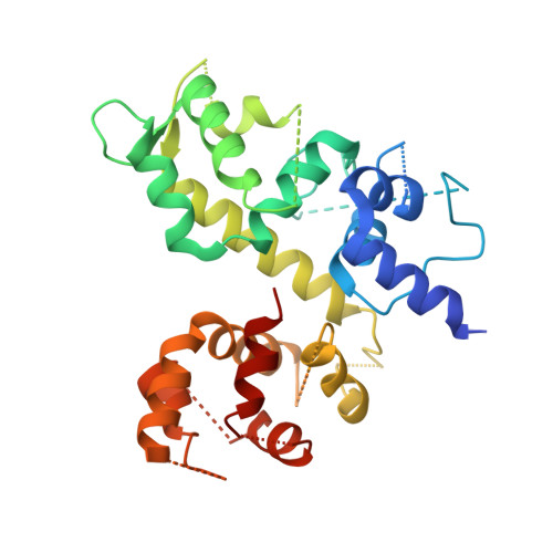
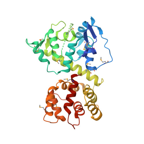
Structure of the MICU1-MICU2 heterodimer provides insights into the gatekeeping threshold shift.

Park, J.Lee, Y.Park, T.Kang, J.Y.Mun, S.A.Jin, M.Yang, J.Eom, S.H.

(2020) IUCrJ 7: 355-365

  • DOI: https://doi.org/10.1107/S2052252520001840
  • Primary Citation of Related Structures:  
    6LE5

  • PubMed Abstract: 

    Mitochondrial calcium uptake proteins 1 and 2 (MICU1 and MICU2) mediate mitochondrial Ca 2+ influx via the mitochondrial calcium uniporter (MCU). Its molecular action for Ca 2+ uptake is tightly controlled by the MICU1-MICU2 heterodimer, which comprises Ca 2+ sensing proteins which act as gatekeepers at low [Ca 2+ ] or facilitators at high [Ca 2+ ]. However, the mechanism underlying the regulation of the Ca 2+ gatekeeping threshold for mitochondrial Ca 2+ uptake through the MCU by the MICU1-MICU2 heterodimer remains unclear. In this study, we determined the crystal structure of the apo form of the human MICU1-MICU2 heterodimer that functions as the MCU gatekeeper. MICU1 and MICU2 assemble in the face-to-face heterodimer with salt bridges and me-thio-nine knobs stabilizing the heterodimer in an apo state. Structural analysis suggests how the heterodimer sets a higher Ca 2+ threshold than the MICU1 homodimer. The structure of the heterodimer in the apo state provides a framework for understanding the gatekeeping role of the MICU1-MICU2 heterodimer.


  • Organizational Affiliation

    School of Life Sciences, Gwangju Institute of Science and Technology (GIST), Buk-gu, Gwangju 61005, Republic of Korea.


Macromolecules
Find similar proteins by:  (by identity cutoff)  |  3D Structure
Entity ID: 1
MoleculeChains Sequence LengthOrganismDetailsImage
Calcium uptake protein 1, mitochondrial
A, D, E, G
348Homo sapiensMutation(s): 0 
Gene Names: MICU1
UniProt & NIH Common Fund Data Resources
Find proteins for Q9BPX6 (Homo sapiens)
Explore Q9BPX6 
Go to UniProtKB:  Q9BPX6
PHAROS:  Q9BPX6
GTEx:  ENSG00000107745 
Entity Groups  
Sequence Clusters30% Identity50% Identity70% Identity90% Identity95% Identity100% Identity
UniProt GroupQ9BPX6
Sequence Annotations
Expand
  • Reference Sequence
Find similar proteins by:  (by identity cutoff)  |  3D Structure
Entity ID: 2
MoleculeChains Sequence LengthOrganismDetailsImage
Calcium uptake protein 2, mitochondrial
B, C, F, H
325Homo sapiensMutation(s): 0 
Gene Names: MICU2
UniProt & NIH Common Fund Data Resources
Find proteins for Q8IYU8 (Homo sapiens)
Explore Q8IYU8 
Go to UniProtKB:  Q8IYU8
PHAROS:  Q8IYU8
GTEx:  ENSG00000165487 
Entity Groups  
Sequence Clusters30% Identity50% Identity70% Identity90% Identity95% Identity100% Identity
UniProt GroupQ8IYU8
Sequence Annotations
Expand
  • Reference Sequence
Small Molecules
Modified Residues  1 Unique
IDChains TypeFormula2D DiagramParent
MSE
Query on MSE
B, C, F, H
L-PEPTIDE LINKINGC5 H11 N O2 SeMET
Experimental Data & Validation

Experimental Data

  • Method: X-RAY DIFFRACTION
  • Resolution: 3.10 Å
  • R-Value Free: 0.332 
  • R-Value Work: 0.293 
  • R-Value Observed: 0.295 
  • Space Group: P 1 21 1
Unit Cell:
Length ( Å )Angle ( ˚ )
a = 62.97α = 90
b = 173.72β = 93.88
c = 148γ = 90
Software Package:
Software NamePurpose
REFMACrefinement
iMOSFLMdata reduction
Cootmodel building
SHELXphasing
Aimlessdata scaling

Structure Validation

View Full Validation Report


Entry History 

Deposition Data

Revision History  (Full details and data files)

  • Version 1.0: 2020-03-04
    Type: Initial release
  • Version 1.1: 2020-03-25
    Changes: Database references