6K9V | pdb_00006k9v

Crystal structure of tubulin in complex with inhibitor D64


Experimental Data Snapshot

  • Method: X-RAY DIFFRACTION
  • Resolution: 2.54 Å
  • R-Value Free: 
    0.248 (Depositor), 0.256 (DCC) 
  • R-Value Work: 
    0.216 (Depositor), 0.226 (DCC) 
  • R-Value Observed: 
    0.217 (Depositor) 

Starting Model: experimental
View more details

wwPDB Validation 3D Report Full Report

Validation slider image for 6K9V

Ligand Structure Quality Assessment 


This is version 1.2 of the entry. See complete history

Literature

Structural insights into the design of indole derivatives as tubulin polymerization inhibitors.

Li, Y.Yang, J.Niu, L.Hu, D.Li, H.Chen, L.Yu, Y.Chen, Q.

(2020) FEBS Lett 594: 199-204

  • DOI: https://doi.org/10.1002/1873-3468.13566
  • Primary Citation Related Structures: 
    6K9V

  • PubMed Abstract: 

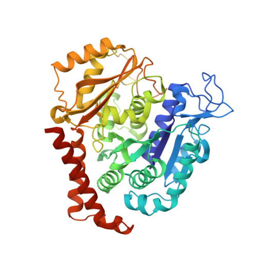
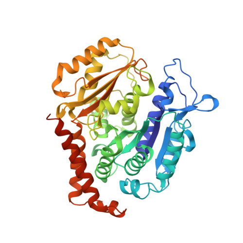
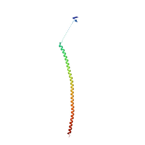
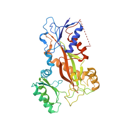
    Microtubules are composed of αβ-tubulin heterodimers, and drugs that interfere with microtubule dynamics are used widely in cancer chemotherapy. Small synthetic molecules with an indole nucleus as a core structure have been identified as microtubule inhibitors and recognized as anticancer agents. However, structural information for the interactions between indole derivatives and tubulin is sparse. Here, we present the 2.55 Å crystal structure of tubulin in complex with the indole derivative D64131. We compare the binding modes of D64131, colchicine, and five other indole derivatives to tubulin. These results reveal the interactions between the indole derivatives and tubulin, explain previous results of structure-activity-relationship (SAR) studies and, thus, provide insights into the development of new indole derivatives targeting the colchicine binding site.


  • Organizational Affiliation
    • Cancer Center, West China Hospital, Sichuan University, and Collaborative Innovation Center of Biotherapy, Chengdu, China.

Macromolecule Content 

  • Total Structure Weight: 264.68 kDa 
  • Atom Count: 17,921 
  • Modeled Residue Count: 2,180 
  • Deposited Residue Count: 2,317 
  • Unique protein chains: 4

Macromolecules

Find similar proteins by:|  3D Structure
Entity ID: 1
MoleculeChains  Sequence LengthOrganismDetailsImage
Tubulin alpha-1B chain
A, C
450Bos taurusMutation(s): 0 
EC: 3.6.5
UniProt
Find proteins for P81947 (Bos taurus)
Explore P81947 
Go to UniProtKB:  P81947
Entity Groups
Sequence Clusters30% Identity50% Identity70% Identity90% Identity95% Identity100% Identity
UniProt GroupP81947
Sequence Annotations
Expand
Reference Sequence
Find similar proteins by:|  3D Structure
Entity ID: 2
MoleculeChains  Sequence LengthOrganismDetailsImage
Tubulin beta-2B chain
B, D
445Bos taurusMutation(s): 0 
UniProt
Find proteins for Q6B856 (Bos taurus)
Explore Q6B856 
Go to UniProtKB:  Q6B856
Entity Groups
Sequence Clusters30% Identity50% Identity70% Identity90% Identity95% Identity100% Identity
UniProt GroupQ6B856
Sequence Annotations
Expand
Reference Sequence
Find similar proteins by:|  3D Structure
Entity ID: 3
MoleculeChains  Sequence LengthOrganismDetailsImage
Stathmin-4143Rattus norvegicusMutation(s): 0 
Gene Names: Stmn4
UniProt
Find proteins for P63043 (Rattus norvegicus)
Explore P63043 
Go to UniProtKB:  P63043
Entity Groups
Sequence Clusters30% Identity50% Identity70% Identity90% Identity95% Identity100% Identity
UniProt GroupP63043
Sequence Annotations
Expand
Reference Sequence
Find similar proteins by:|  3D Structure
Entity ID: 4
MoleculeChains  Sequence LengthOrganismDetailsImage
Tubulin tyrosine ligase384Gallus gallusMutation(s): 0 
Gene Names: TTL

Small Molecules

Ligands 7 Unique
IDChains Name / Formula / InChI Key2D Diagram3D Interactions
GTP

Query on GTP


Download:Ideal Coordinates CCD File
G [auth A],
N [auth C],
Q [auth D]
GUANOSINE-5'-TRIPHOSPHATE
C10 H16 N5 O14 P3
XKMLYUALXHKNFT-UUOKFMHZSA-N
ACP

Query on ACP


Download:Ideal Coordinates CCD File
T [auth F]PHOSPHOMETHYLPHOSPHONIC ACID ADENYLATE ESTER
C11 H18 N5 O12 P3
UFZTZBNSLXELAL-IOSLPCCCSA-N
GDP

Query on GDP


Download:Ideal Coordinates CCD File
J [auth B]GUANOSINE-5'-DIPHOSPHATE
C10 H15 N5 O11 P2
QGWNDRXFNXRZMB-UUOKFMHZSA-N
D3L
(Subject of Investigation/LOI)

Query on D3L


Download:Ideal Coordinates CCD File
M [auth B],
R [auth D]
(5-methoxy-1H-indol-2-yl)-phenyl-methanone
C16 H13 N O2
ICMIJSRDISNKOC-UHFFFAOYSA-N
MES

Query on MES


Download:Ideal Coordinates CCD File
L [auth B]2-(N-MORPHOLINO)-ETHANESULFONIC ACID
C6 H13 N O4 S
SXGZJKUKBWWHRA-UHFFFAOYSA-N
CA

Query on CA


Download:Ideal Coordinates CCD File
P [auth C]CALCIUM ION
Ca
BHPQYMZQTOCNFJ-UHFFFAOYSA-N
MG

Query on MG


Download:Ideal Coordinates CCD File
H [auth A],
I [auth A],
K [auth B],
O [auth C],
S [auth D]
MAGNESIUM ION
Mg
JLVVSXFLKOJNIY-UHFFFAOYSA-N
Binding Affinity Annotations 
IDSourceBinding Affinity
D3L BindingDB:  6K9V IC50: 530 (nM) from 1 assay(s)

Experimental Data & Validation

Experimental Data

  • Method: X-RAY DIFFRACTION
  • Resolution: 2.54 Å
  • R-Value Free:  0.248 (Depositor), 0.256 (DCC) 
  • R-Value Work:  0.216 (Depositor), 0.226 (DCC) 
  • R-Value Observed: 0.217 (Depositor) 
Space Group: P 21 21 21
Unit Cell:
Length ( Å )Angle ( ˚ )
a = 104.219α = 90
b = 157.435β = 90
c = 180.045γ = 90
Software Package:
Software NamePurpose
PHENIXrefinement
HKL-3000data reduction
HKL-3000data scaling
PHASERphasing

Structure Validation

View Full Validation Report


Ligand Structure Quality Assessment 


Entry History 

Deposition Data

Revision History  (Full details and data files)

  • Version 1.0: 2019-08-28
    Type: Initial release
  • Version 1.1: 2020-01-22
    Changes: Database references
  • Version 1.2: 2023-11-22
    Changes: Data collection, Database references, Derived calculations, Refinement description