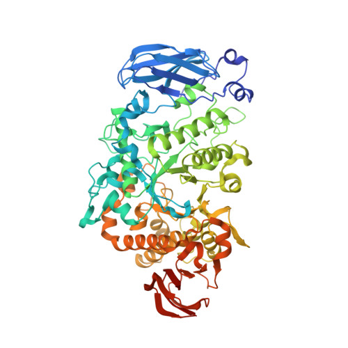
6JFJ

Crystal structure of Pullulanase from Paenibacillus barengoltzii complex with maltohexaose and alpha-cyclodextrin


Experimental Data Snapshot

  • Method: X-RAY DIFFRACTION
  • Resolution: 1.93 Å
  • R-Value Free: 0.189 
  • R-Value Work: 0.145 
  • R-Value Observed: 0.148 

wwPDB Validation   3D Report Full Report


This is version 2.0 of the entry. See complete history


Literature

Structural basis of carbohydrate binding in domain C of a type I pullulanase from Paenibacillus barengoltzii.

Huang, P.Wu, S.Yang, S.Yan, Q.Jiang, Z.

(2020) Acta Crystallogr D Struct Biol 76: 447-457

  • DOI: https://doi.org/10.1107/S205979832000409X
  • Primary Citation of Related Structures:  
    6JEQ, 6JFJ, 6JFX, 6JHF

  • PubMed Abstract: 
  • Pullulanase (EC 3.2.1.41) is a well known starch-debranching enzyme that catalyzes the cleavage of α-1,6-glycosidic linkages in α-glucans such as starch and pullulan. Crystal structures of a type I pullulanase from Paenibacillus barengoltzii (PbPulA) and of PbPulA in complex with maltopentaose (G5), maltohexaose (G6)/α-cyclodextrin (α-CD) and β-cyclodextrin (β-CD) were determined in order to better understand substrate binding to this enzyme ...

    Pullulanase (EC 3.2.1.41) is a well known starch-debranching enzyme that catalyzes the cleavage of α-1,6-glycosidic linkages in α-glucans such as starch and pullulan. Crystal structures of a type I pullulanase from Paenibacillus barengoltzii (PbPulA) and of PbPulA in complex with maltopentaose (G5), maltohexaose (G6)/α-cyclodextrin (α-CD) and β-cyclodextrin (β-CD) were determined in order to better understand substrate binding to this enzyme. PbPulA belongs to glycoside hydrolase (GH) family 13 subfamily 14 and is composed of three domains (CBM48, A and C). Three carbohydrate-binding sites identified in PbPulA were located in CBM48, near the active site and in domain C, respectively. The binding site in CBM48 was specific for β-CD, while that in domain C has not been reported for other pullulanases. The domain C binding site had higher affinity for α-CD than for G6; a small motif (FGGEH) seemed to be one of the major determinants for carbohydrate binding in this domain. Structure-based mutations of several surface-exposed aromatic residues in CBM48 and domain C had a debilitating effect on the activity of the enzyme. These results suggest that both CBM48 and domain C play a role in binding substrates. The crystal forms described contribute to the understanding of pullulanase domain-carbohydrate interactions.


    Organizational Affiliation

    Beijing Advanced Innovation Center for Food Nutrition and Human Health, College of Food Science and Nutritional Engineering, China Agricultural University, Beijing 100083, People's Republic of China.


Macromolecules
Find similar proteins by:  (by identity cutoff)  |  3D Structure
Entity ID: 1
MoleculeChainsSequence LengthOrganismDetailsImage
Pulullanase675Paenibacillus barengoltziiMutation(s): 0 
EC: 3.2.1.41
UniProt
Find proteins for A0A0C5GWS2 (Paenibacillus barengoltzii)
Explore A0A0C5GWS2 
Go to UniProtKB:  A0A0C5GWS2
Entity Groups  
Sequence Clusters30% Identity50% Identity70% Identity90% Identity95% Identity100% Identity
UniProt GroupA0A0C5GWS2
Protein Feature View
Expand
  • Reference Sequence
Oligosaccharides

Help

Entity ID: 2
MoleculeChainsChain Length2D DiagramGlycosylation3D Interactions
alpha-D-glucopyranose-(1-4)-alpha-D-glucopyranose-(1-4)-alpha-D-glucopyranose
B, C
3N/A Oligosaccharides Interaction
Glycosylation Resources
GlyTouCan:  G96370VA
GlyCosmos:  G96370VA
GlyGen:  G96370VA
Entity ID: 3
MoleculeChainsChain Length2D DiagramGlycosylation3D Interactions
alpha-D-glucopyranose-(1-4)-alpha-D-glucopyranose-(1-4)-alpha-D-glucopyranose-(1-4)-alpha-D-glucopyranose-(1-4)-alpha-D-glucopyranose
D
5N/A Oligosaccharides Interaction
Glycosylation Resources
GlyTouCan:  G50146AM
GlyCosmos:  G50146AM
Small Molecules
Ligands 8 Unique
IDChainsName / Formula / InChI Key2D Diagram3D Interactions
1PE
Query on 1PE

Download Ideal Coordinates CCD File 
U [auth A]PENTAETHYLENE GLYCOL
C10 H22 O6
JLFNLZLINWHATN-UHFFFAOYSA-N
 Ligand Interaction
TAM
Query on TAM

Download Ideal Coordinates CCD File 
V [auth A],
W [auth A]
TRIS(HYDROXYETHYL)AMINOMETHANE
C7 H17 N O3
GKODZWOPPOTFGA-UHFFFAOYSA-N
 Ligand Interaction
PGE
Query on PGE

Download Ideal Coordinates CCD File 
S [auth A],
T [auth A]
TRIETHYLENE GLYCOL
C6 H14 O4
ZIBGPFATKBEMQZ-UHFFFAOYSA-N
 Ligand Interaction
PEG
Query on PEG

Download Ideal Coordinates CCD File 
I [auth A]
J [auth A]
K [auth A]
L [auth A]
M [auth A]
I [auth A],
J [auth A],
K [auth A],
L [auth A],
M [auth A],
N [auth A],
O [auth A],
P [auth A],
Q [auth A],
R [auth A]
DI(HYDROXYETHYL)ETHER
C4 H10 O3
MTHSVFCYNBDYFN-UHFFFAOYSA-N
 Ligand Interaction
GOL
Query on GOL

Download Ideal Coordinates CCD File 
E [auth A],
F [auth A],
G [auth A],
H [auth A]
GLYCEROL
C3 H8 O3
PEDCQBHIVMGVHV-UHFFFAOYSA-N
 Ligand Interaction
IMD
Query on IMD

Download Ideal Coordinates CCD File 
AA [auth A],
X [auth A],
Y [auth A],
Z [auth A]
IMIDAZOLE
C3 H5 N2
RAXXELZNTBOGNW-UHFFFAOYSA-O
 Ligand Interaction
CA
Query on CA

Download Ideal Coordinates CCD File 
BA [auth A],
CA [auth A]
CALCIUM ION
Ca
BHPQYMZQTOCNFJ-UHFFFAOYSA-N
 Ligand Interaction
CL
Query on CL

Download Ideal Coordinates CCD File 
DA [auth A]
EA [auth A]
FA [auth A]
GA [auth A]
HA [auth A]
DA [auth A],
EA [auth A],
FA [auth A],
GA [auth A],
HA [auth A],
IA [auth A],
JA [auth A],
KA [auth A]
CHLORIDE ION
Cl
VEXZGXHMUGYJMC-UHFFFAOYSA-M
 Ligand Interaction
Biologically Interesting Molecules (External Reference) 2 Unique
Entity ID: 2
IDChainsNameType/Class2D Diagram3D Interactions
PRD_900009
Query on PRD_900009
B, C
alpha-maltotrioseOligosaccharide / Nutrient Ligand Interaction
Entity ID: 3
IDChainsNameType/Class2D Diagram3D Interactions
PRD_900030
Query on PRD_900030
D
alpha-maltopentaoseOligosaccharide / Substrate analog Ligand Interaction
Experimental Data & Validation

Experimental Data

  • Method: X-RAY DIFFRACTION
  • Resolution: 1.93 Å
  • R-Value Free: 0.189 
  • R-Value Work: 0.145 
  • R-Value Observed: 0.148 
  • Space Group: P 2 21 21
Unit Cell:
Length ( Å )Angle ( ˚ )
a = 49.911α = 90
b = 98.179β = 90
c = 140.187γ = 90
Software Package:
Software NamePurpose
PHENIXrefinement
HKL-2000data reduction
HKL-2000data scaling
PHENIXphasing

Structure Validation

View Full Validation Report


Entry History 

Deposition Data

Revision History  (Full details and data files)

  • Version 1.0: 2019-02-20
    Type: Initial release
  • Version 1.1: 2020-05-06
    Changes: Data collection, Database references
  • Version 1.2: 2020-05-13
    Changes: Database references
  • Version 2.0: 2020-07-29
    Type: Remediation
    Reason: Carbohydrate remediation
    Changes: Atomic model, Data collection, Derived calculations, Structure summary