Structural basis of carbohydrate binding in domain C of a type I pullulanase from Paenibacillus barengoltzii.
Huang, P., Wu, S., Yang, S., Yan, Q., Jiang, Z.(2020) Acta Crystallogr D Struct Biol 76: 447-457
- PubMed: 32355041 Search on PubMed
- DOI: https://doi.org/10.1107/S205979832000409X
- Primary Citation Related Structures:
6JEQ, 6JFJ, 6JFX, 6JHF - PubMed Abstract:
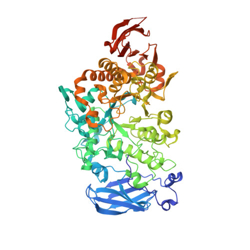
Pullulanase (EC 3.2.1.41) is a well known starch-debranching enzyme that catalyzes the cleavage of α-1,6-glycosidic linkages in α-glucans such as starch and pullulan. Crystal structures of a type I pullulanase from Paenibacillus barengoltzii (PbPulA) and of PbPulA in complex with maltopentaose (G5), maltohexaose (G6)/α-cyclodextrin (α-CD) and β-cyclodextrin (β-CD) were determined in order to better understand substrate binding to this enzyme. PbPulA belongs to glycoside hydrolase (GH) family 13 subfamily 14 and is composed of three domains (CBM48, A and C). Three carbohydrate-binding sites identified in PbPulA were located in CBM48, near the active site and in domain C, respectively. The binding site in CBM48 was specific for β-CD, while that in domain C has not been reported for other pullulanases. The domain C binding site had higher affinity for α-CD than for G6; a small motif (FGGEH) seemed to be one of the major determinants for carbohydrate binding in this domain. Structure-based mutations of several surface-exposed aromatic residues in CBM48 and domain C had a debilitating effect on the activity of the enzyme. These results suggest that both CBM48 and domain C play a role in binding substrates. The crystal forms described contribute to the understanding of pullulanase domain-carbohydrate interactions.
- Beijing Advanced Innovation Center for Food Nutrition and Human Health, College of Food Science and Nutritional Engineering, China Agricultural University, Beijing 100083, People's Republic of China.
Organizational Affiliation: