6IDX

Crystal Structure of BAI1/ELMO2 complex


Experimental Data Snapshot

  • Method: X-RAY DIFFRACTION
  • Resolution: 1.70 Å
  • R-Value Free: 0.206 
  • R-Value Work: 0.179 
  • R-Value Observed: 0.181 

wwPDB Validation   3D Report Full Report


This is version 1.0 of the entry. See complete history


Literature

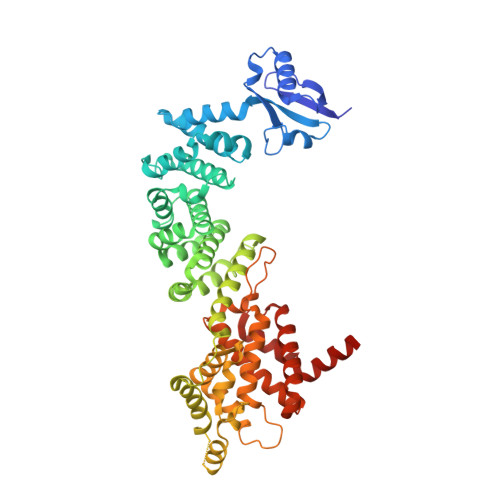
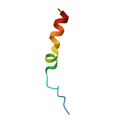
Structure of BAI1/ELMO2 complex reveals an action mechanism of adhesion GPCRs via ELMO family scaffolds

Weng, Z.Situ, C.Lin, L.Wu, Z.Zhu, J.Zhang, R.

(2019) Nat Commun 10: 51-51

  • DOI: https://doi.org/10.1038/s41467-018-07938-9
  • Primary Citation of Related Structures:  
    6IDX, 6IE1

  • PubMed Abstract: 
  • The brain-specific angiogenesis inhibitor (BAI) subfamily of adhesion G protein-coupled receptors (aGPCRs) plays crucial roles in diverse cellular processes including phagocytosis, myoblast fusion, and synaptic development through the ELMO/DOCK/Rac signaling pathway, although the underlying molecular mechanism is not well understood ...

    The brain-specific angiogenesis inhibitor (BAI) subfamily of adhesion G protein-coupled receptors (aGPCRs) plays crucial roles in diverse cellular processes including phagocytosis, myoblast fusion, and synaptic development through the ELMO/DOCK/Rac signaling pathway, although the underlying molecular mechanism is not well understood. Here, we demonstrate that an evolutionarily conserved fragment located in the C-terminal cytoplasmic tail of BAI-aGPCRs is specifically recognized by the RBD-ARR-ELMO (RAE) supramodule of the ELMO family scaffolds. The crystal structures of ELMO2-RAE and its complex with BAI1 uncover the molecular basis of BAI/ELMO interactions. Based on the complex structure we identify aGPCR-GPR128 as another upstream receptor for the ELMO family scaffolds, most likely with a recognition mode similar to that of BAI/ELMO interactions. Finally, we map disease-causing mutations of BAI and ELMO and analyze their effects on complex formation.


    Organizational Affiliation

    School of Life Science and Technology, ShanghaiTech University, 100 Haike Road, 201210, Shanghai, China. rgzhang@sibcb.ac.cn.


Macromolecules
Find similar proteins by:  (by identity cutoff)  |  3D Structure
Entity ID: 1
MoleculeChainsSequence LengthOrganismDetailsImage
Engulfment and cell motility protein 2524Homo sapiensMutation(s): 0 
Gene Names: ELMO2CED12AKIAA1834
UniProt & NIH Common Fund Data Resources
Find proteins for Q96JJ3 (Homo sapiens)
Explore Q96JJ3 
Go to UniProtKB:  Q96JJ3
PHAROS:  Q96JJ3
Entity Groups  
Sequence Clusters30% Identity50% Identity70% Identity90% Identity95% Identity100% Identity
UniProt GroupQ96JJ3
Protein Feature View
Expand
  • Reference Sequence

Find similar proteins by:  Sequence   |   3D Structure  

Entity ID: 2
MoleculeChainsSequence LengthOrganismDetailsImage
Adhesion G protein-coupled receptor B1B [auth C]25Mus musculusMutation(s): 0 
Gene Names: Adgrb1Bai1
UniProt & NIH Common Fund Data Resources
Find proteins for Q3UHD1 (Mus musculus)
Explore Q3UHD1 
Go to UniProtKB:  Q3UHD1
IMPC:  MGI:1933736
Entity Groups  
Sequence Clusters30% Identity50% Identity70% Identity90% Identity95% Identity100% Identity
UniProt GroupQ3UHD1
Protein Feature View
Expand
  • Reference Sequence
Experimental Data & Validation

Experimental Data

  • Method: X-RAY DIFFRACTION
  • Resolution: 1.70 Å
  • R-Value Free: 0.206 
  • R-Value Work: 0.179 
  • R-Value Observed: 0.181 
  • Space Group: P 31 2 1
Unit Cell:
Length ( Å )Angle ( ˚ )
a = 92.869α = 90
b = 92.869β = 90
c = 130.762γ = 120
Software Package:
Software NamePurpose
PHENIXrefinement
XSCALEdata scaling
PDB_EXTRACTdata extraction
d*TREKdata reduction
PHASERphasing

Structure Validation

View Full Validation Report


Entry History 

Deposition Data

Revision History  (Full details and data files)

  • Version 1.0: 2019-01-23
    Type: Initial release