6GXC | pdb_00006gxc

Bacterial oligosaccharyltransferase PglB in complex with an inhibitory peptide and a reactive lipid-linked oligosaccharide analog


Experimental Data Snapshot

  • Method: X-RAY DIFFRACTION
  • Resolution: 3.40 Å
  • R-Value Free: 
    0.283 (Depositor), 0.285 (DCC) 
  • R-Value Work: 
    0.249 (Depositor), 0.256 (DCC) 
  • R-Value Observed: 
    0.252 (Depositor) 

wwPDB Validation 3D Report Full Report

Validation slider image for 6GXC

Ligand Structure Quality Assessment 


This is version 1.1 of the entry. See complete history

Literature

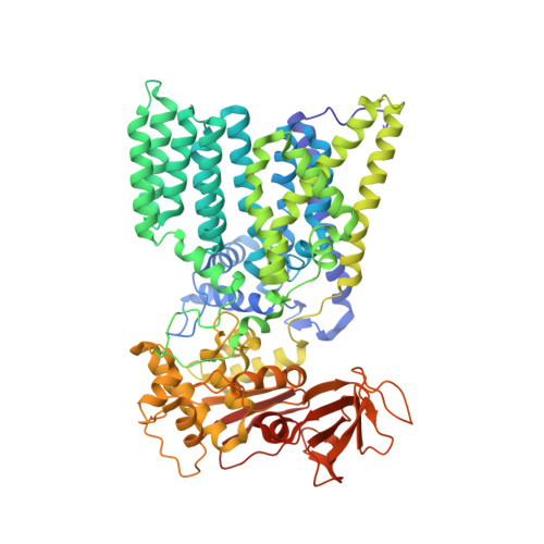
Structure of bacterial oligosaccharyltransferase PglB bound to a reactive LLO and an inhibitory peptide.

Napiorkowska, M.Boilevin, J.Darbre, T.Reymond, J.L.Locher, K.P.

(2018) Sci Rep 8: 16297-16297

  • DOI: https://doi.org/10.1038/s41598-018-34534-0
  • Primary Citation Related Structures: 
    6GXC

  • PubMed Abstract: 

    Oligosaccharyltransferase (OST) is a key enzyme of the N-glycosylation pathway, where it catalyzes the transfer of a glycan from a lipid-linked oligosaccharide (LLO) to an acceptor asparagine within the conserved sequon N-X-T/S. A previous structure of a ternary complex of bacterial single subunit OST, PglB, bound to a non-hydrolyzable LLO analog and a wild type acceptor peptide showed how both substrates bind and how an external loop (EL5) of the enzyme provided specific substrate-binding contacts. However, there was a relatively large separation of the substrates at the active site. Here we present the X-ray structure of PglB bound to a reactive LLO analog and an inhibitory peptide, revealing previously unobserved interactions in the active site. We found that the atoms forming the N-glycosidic bond (C-1 of the GlcNAc moiety of LLO and the -NH 2 group of the peptide) are closer than in the previous structure, suggesting that we have captured a conformation closer to the transition state of the reaction. We find that the distance between the divalent metal ion and the glycosidic oxygen of LLO is now 4 Å, suggesting that the metal stabilizes the leaving group of the nucleophilic substitution reaction. Further, the carboxylate group of a conserved aspartate of PglB mediates an interaction network between the reducing-end sugar of the LLO, the asparagine side chain of the acceptor peptide, and a bound divalent metal ion. The interactions identified in this novel state are likely to be relevant in the catalytic mechanisms of all OSTs.


  • Organizational Affiliation
    • Institute of Molecular Biology and Biophysics, ETH Zurich, Zurich, Switzerland.

Macromolecule Content 

  • Total Structure Weight: 86.49 kDa 
  • Atom Count: 5,931 
  • Modeled Residue Count: 714 
  • Deposited Residue Count: 732 
  • Unique protein chains: 2

Macromolecules

Find similar proteins by:|  3D Structure
Entity ID: 1
MoleculeChains  Sequence LengthOrganismDetailsImage
Undecaprenyl-diphosphooligosaccharide--protein glycotransferase724Campylobacter lari RM2100Mutation(s): 24 
Gene Names: pglBCla_1253
EC: 2.4.99.19
Membrane Entity: Yes 
UniProt
Find proteins for B9KDD4 (Campylobacter lari (strain RM2100 / D67 / ATCC BAA-1060))
Explore B9KDD4 
Go to UniProtKB:  B9KDD4
Entity Groups
Sequence Clusters30% Identity50% Identity70% Identity90% Identity95% Identity100% Identity
UniProt GroupB9KDD4
Sequence Annotations
Expand
Reference Sequence
Find similar proteins by:  Sequence   |   3D Structure  
Entity ID: 2
MoleculeChains  Sequence LengthOrganismDetailsImage
GLY-ASP-GLN-DAB-ALA-THR-PPN-GLY8Campylobacter lari RM2100Mutation(s): 0 

Small Molecules

Ligands 5 Unique
IDChains Name / Formula / InChI Key2D Diagram3D Interactions
FFK

Query on FFK


Download:Ideal Coordinates CCD File
H [auth A][(2~{R},3~{R},4~{R},5~{S},6~{R})-3-acetamido-6-(hydroxymethyl)-4,5-bis(oxidanyl)oxan-2-yl] [oxidanyl-[(2~{Z},6~{Z},10~{Z})-3,7,11,15-tetramethylhexadeca-2,6,10,14-tetraenoxy]phosphoryl] hydrogen phosphate
C28 H49 N O12 P2
MCKZOQNLWLVDRX-ADLZVGHSSA-N
MES

Query on MES


Download:Ideal Coordinates CCD File
F [auth A]2-(N-MORPHOLINO)-ETHANESULFONIC ACID
C6 H13 N O4 S
SXGZJKUKBWWHRA-UHFFFAOYSA-N
PEG

Query on PEG


Download:Ideal Coordinates CCD File
G [auth A]DI(HYDROXYETHYL)ETHER
C4 H10 O3
MTHSVFCYNBDYFN-UHFFFAOYSA-N
MN

Query on MN


Download:Ideal Coordinates CCD File
C [auth A],
D [auth A]
MANGANESE (II) ION
Mn
WAEMQWOKJMHJLA-UHFFFAOYSA-N
NA

Query on NA


Download:Ideal Coordinates CCD File
E [auth A]SODIUM ION
Na
FKNQFGJONOIPTF-UHFFFAOYSA-N
Modified Residues  2 Unique
IDChains TypeFormula2D DiagramParent
DAB
Query on DAB
B
L-PEPTIDE LINKINGC4 H10 N2 O2ALA
PPN
Query on PPN
B
L-PEPTIDE LINKINGC9 H10 N2 O4PHE

Experimental Data & Validation

Experimental Data

  • Method: X-RAY DIFFRACTION
  • Resolution: 3.40 Å
  • R-Value Free:  0.283 (Depositor), 0.285 (DCC) 
  • R-Value Work:  0.249 (Depositor), 0.256 (DCC) 
  • R-Value Observed: 0.252 (Depositor) 
Space Group: P 21 21 21
Unit Cell:
Length ( Å )Angle ( ˚ )
a = 83.81α = 90
b = 116.54β = 90
c = 173.89γ = 90
Software Package:
Software NamePurpose
PHENIXrefinement
XDSdata reduction
XSCALEdata scaling
PHENIXphasing

Structure Validation

View Full Validation Report


Ligand Structure Quality Assessment 


Entry History 

& Funding Information

Deposition Data


Funding OrganizationLocationGrant Number
Swiss National Science FoundationSwitzerlandSNF 310030B_166672

Revision History  (Full details and data files)

  • Version 1.0: 2018-11-14
    Type: Initial release
  • Version 1.1: 2025-04-09
    Changes: Advisory, Data collection, Database references, Derived calculations, Structure summary