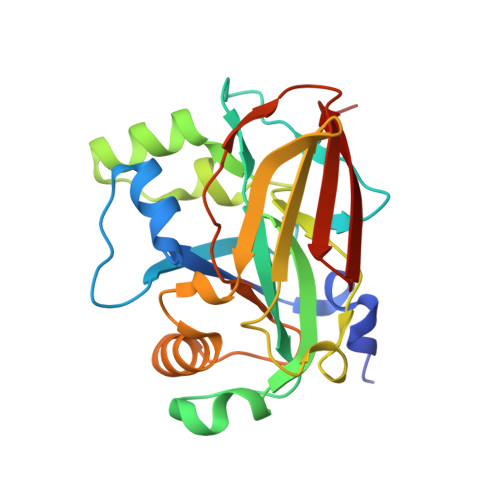
6FOG

X-ray structure of homo sapiens Fumarylacetoacetate hydrolase domain containing protein 1 (FAHD1) in complex with inhibitor oxalate at 1.94A resolution.

  • Classification: hydrolase/lyase
  • Organism(s): Homo sapiens
  • Expression System: Escherichia coli
  • Mutation(s): No 

  • Deposited: 2018-02-07 Released: 2018-11-07 
  • Deposition Author(s): Naschberger, A., Weiss, A.K.H.
  • Funding Organization(s): European integrated FP6-LIFESCIHEALTH project MiMAGE, Austria Wirtschaftsservice Gesellschaft (AWS), Austrian Science Fund

Experimental Data Snapshot

  • Method: X-RAY DIFFRACTION
  • Resolution: 1.94 Å
  • R-Value Free: 0.208 
  • R-Value Work: 0.181 
  • R-Value Observed: 0.182 

wwPDB Validation   3D Report Full Report


Ligand Structure Quality Assessment 


This is version 1.2 of the entry. See complete history


Literature

Structural basis for the bi-functionality of human oxaloacetate decarboxylase FAHD1.

Weiss, A.K.H.Naschberger, A.Loeffler, J.R.Gstach, H.Bowler, M.W.Holzknecht, M.Cappuccio, E.Pittl, A.Etemad, S.Dunzendorfer-Matt, T.Scheffzek, K.Liedl, K.R.Jansen-Durr, P.

(2018) Biochem J 475: 3561-3576

  • DOI: https://doi.org/10.1042/BCJ20180750
  • Primary Citation of Related Structures:  
    6FOG, 6FOH

  • PubMed Abstract: 

    Whereas enzymes in the fumarylacetoacetate hydrolase (FAH) superfamily catalyze several distinct chemical reactions, the structural basis for their multi-functionality remains elusive. As a well-studied example, human FAH domain-containing protein 1 (FAHD1) is a mitochondrial protein displaying both acylpyruvate hydrolase (ApH) and oxaloacetate decarboxylase (ODx) activity. As mitochondrial ODx, FAHD1 acts antagonistically to pyruvate carboxylase, a key metabolic enzyme. Despite its importance for mitochondrial function, very little is known about the catalytic mechanisms underlying FAHD1 enzymatic activities, and the architecture of its ligated active site is currently ill defined. We present crystallographic data of human FAHD1 that provide new insights into the structure of the catalytic center at high resolution, featuring a flexible 'lid'-like helical region which folds into a helical structure upon binding of the ODx inhibitor oxalate. The oxalate-driven structural transition results in the generation of a potential catalytic triad consisting of E33, H30 and an associated water molecule. In silico docking studies indicate that the substrate is further stabilized by a complex hydrogen-bond network, involving amino acids Q109 and K123, identified herein as potential key residues for FAHD1 catalytic activity. Mutation of amino acids H30, E33 and K123 each had discernible influence on the ApH and/or ODx activity of FAHD1, suggesting distinct catalytic mechanisms for both activities. The structural analysis presented here provides a defined structural map of the active site of FAHD1 and contributes to a better understanding of the FAH superfamily of enzymes.


  • Organizational Affiliation

    Research Institute for Biomedical Aging Research, University of Innsbruck, Rennweg 10, Innsbruck A-6020, Austria.


Macromolecules
Find similar proteins by:  (by identity cutoff)  |  3D Structure
Entity ID: 1
MoleculeChains Sequence LengthOrganismDetailsImage
Acylpyruvase FAHD1, mitochondrial224Homo sapiensMutation(s): 0 
Gene Names: FAHD1C16orf36YISKL
EC: 3.7.1.5 (PDB Primary Data), 4.1.1.3 (PDB Primary Data)
UniProt & NIH Common Fund Data Resources
Find proteins for Q6P587 (Homo sapiens)
Explore Q6P587 
Go to UniProtKB:  Q6P587
PHAROS:  Q6P587
GTEx:  ENSG00000180185 
Entity Groups  
Sequence Clusters30% Identity50% Identity70% Identity90% Identity95% Identity100% Identity
UniProt GroupQ6P587
Sequence Annotations
Expand
  • Reference Sequence
Small Molecules
Ligands 3 Unique
IDChains Name / Formula / InChI Key2D Diagram3D Interactions
OXL (Subject of Investigation/LOI)
Query on OXL

Download Ideal Coordinates CCD File 
AA [auth C]
CA [auth H]
EA [auth E]
J [auth A]
N [auth G]
AA [auth C],
CA [auth H],
EA [auth E],
J [auth A],
N [auth G],
R [auth F],
V [auth D],
X [auth B]
OXALATE ION
C2 O4
MUBZPKHOEPUJKR-UHFFFAOYSA-L
CL
Query on CL

Download Ideal Coordinates CCD File 
FA [auth E],
K [auth A],
S [auth F],
Y [auth B]
CHLORIDE ION
Cl
VEXZGXHMUGYJMC-UHFFFAOYSA-M
MG
Query on MG

Download Ideal Coordinates CCD File 
BA [auth H]
DA [auth E]
I [auth A]
L [auth A]
M [auth G]
BA [auth H],
DA [auth E],
I [auth A],
L [auth A],
M [auth G],
O [auth G],
P [auth F],
Q [auth F],
T [auth D],
U [auth D],
W [auth B],
Z [auth C]
MAGNESIUM ION
Mg
JLVVSXFLKOJNIY-UHFFFAOYSA-N
Experimental Data & Validation

Experimental Data

  • Method: X-RAY DIFFRACTION
  • Resolution: 1.94 Å
  • R-Value Free: 0.208 
  • R-Value Work: 0.181 
  • R-Value Observed: 0.182 
  • Space Group: P 1 21 1
Unit Cell:
Length ( Å )Angle ( ˚ )
a = 75.984α = 90
b = 116.679β = 89.94
c = 125.753γ = 90
Software Package:
Software NamePurpose
REFMACrefinement
XDSdata reduction
XSCALEdata scaling
MOLREPphasing

Structure Validation

View Full Validation Report


Ligand Structure Quality Assessment 


Entry History & Funding Information

Deposition Data


Funding OrganizationLocationGrant Number
European integrated FP6-LIFESCIHEALTH project MiMAGE512020
Austria Wirtschaftsservice Gesellschaft (AWS)Austria--
Austrian Science FundAustriaP28975

Revision History  (Full details and data files)

  • Version 1.0: 2018-11-07
    Type: Initial release
  • Version 1.1: 2018-11-28
    Changes: Data collection, Database references
  • Version 1.2: 2024-05-08
    Changes: Data collection, Database references, Derived calculations, Refinement description