6DKS | pdb_00006dks

Structure of the Rbpj-SHARP-DNA Repressor Complex


Experimental Data Snapshot

  • Method: X-RAY DIFFRACTION
  • Resolution: 2.78 Å
  • R-Value Free: 
    0.228 (Depositor), 0.242 (DCC) 
  • R-Value Work: 
    0.195 (Depositor), 0.206 (DCC) 
  • R-Value Observed: 
    0.197 (Depositor) 

Starting Models: experimental
View more details

wwPDB Validation 3D Report Full Report

Validation slider image for 6DKS

This is version 2.1 of the entry. See complete history

Literature

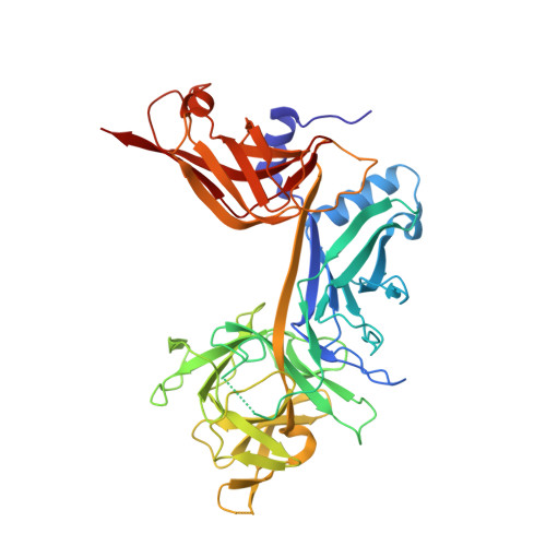
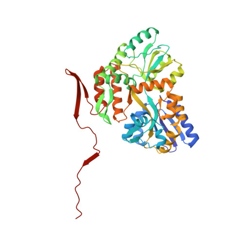
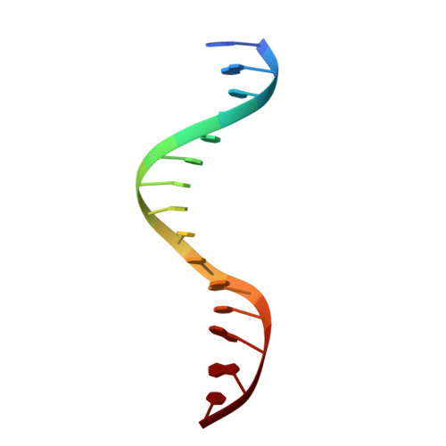
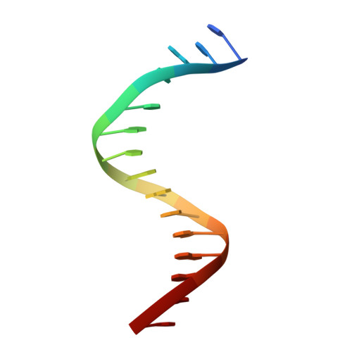
Structural and Functional Studies of the RBPJ-SHARP Complex Reveal a Conserved Corepressor Binding Site.

Yuan, Z.VanderWielen, B.D.Giaimo, B.D.Pan, L.Collins, C.E.Turkiewicz, A.Hein, K.Oswald, F.Borggrefe, T.Kovall, R.A.

(2019) Cell Rep 26: 845-854.e6

  • DOI: https://doi.org/10.1016/j.celrep.2018.12.097
  • Primary Citation Related Structures: 
    6DKS

  • PubMed Abstract: 

    Notch is a conserved signaling pathway that is essential for metazoan development and homeostasis; dysregulated signaling underlies the pathophysiology of numerous human diseases. Receptor-ligand interactions result in gene expression changes, which are regulated by the transcription factor RBPJ. RBPJ forms a complex with the intracellular domain of the Notch receptor and the coactivator Mastermind to activate transcription, but it can also function as a repressor by interacting with corepressor proteins. Here, we determine the structure of RBPJ bound to the corepressor SHARP and DNA, revealing its mode of binding to RBPJ. We tested structure-based mutants in biophysical and biochemical-cellular assays to characterize the role of RBPJ as a repressor, clearly demonstrating that RBPJ mutants deficient for SHARP binding are incapable of repressing transcription of genes responsive to Notch signaling in cells. Altogether, our structure-function studies provide significant insights into the repressor function of RBPJ.


  • Organizational Affiliation
    • Department of Molecular Genetics, Biochemistry and Microbiology, University of Cincinnati College of Medicine, Cincinnati, OH, USA.

Macromolecule Content 

  • Total Structure Weight: 203.47 kDa 
  • Atom Count: 13,438 
  • Modeled Residue Count: 1,711 
  • Deposited Residue Count: 1,718 
  • Unique protein chains: 2
  • Unique nucleic acid chains: 2

Macromolecules


Find similar proteins by:|  3D Structure
Entity ID: 3
MoleculeChains  Sequence LengthOrganismDetailsImage
Recombining binding protein suppressor of hairless
C, G
422Mus musculusMutation(s): 0 
Gene Names: RbpjIgkjrb1IgkrsbpRbpsuh
UniProt & NIH Common Fund Data Resources
Find proteins for P31266 (Mus musculus)
Explore P31266 
Go to UniProtKB:  P31266
IMPC:  MGI:96522
Entity Groups
Sequence Clusters30% Identity50% Identity70% Identity90% Identity95% Identity100% Identity
UniProt GroupP31266
Sequence Annotations
Expand
Reference Sequence
Find similar proteins by:|  3D Structure
Entity ID: 4
MoleculeChains  Sequence LengthOrganismDetailsImage
Maltose/maltodextrin-binding periplasmic protein
D, H
407Escherichia coli O157:H7Mutation(s): 0 
Gene Names: malEZ5632ECs5017
UniProt
Find proteins for P0AEX9 (Escherichia coli (strain K12))
Explore P0AEX9 
Go to UniProtKB:  P0AEX9
Entity Groups
Sequence Clusters30% Identity50% Identity70% Identity90% Identity95% Identity100% Identity
UniProt GroupP0AEX9
Sequence Annotations
Expand
Reference Sequence
Find similar nucleic acids by:  Sequence
Entity ID: 1
MoleculeChains LengthOrganismImage
DNA (5'-D(*AP*AP*TP*CP*TP*TP*TP*CP*CP*CP*AP*CP*AP*GP*T)-3')
A, E
15Mus musculus
Sequence Annotations
Expand
Reference Sequence
Find similar nucleic acids by:  Sequence
Entity ID: 2
MoleculeChains LengthOrganismImage
DNA (5'-D(*TP*TP*AP*CP*TP*GP*TP*GP*GP*GP*AP*AP*AP*GP*A)-3')
B, F
15Mus musculus
Sequence Annotations
Expand
Reference Sequence

Oligosaccharides

Help  
Entity ID: 5
MoleculeChains Length2D Diagram GlycosylationD Interactions
alpha-D-glucopyranose-(1-4)-alpha-D-glucopyranose
I, J
2N/A
Glycosylation Resources
GlyTouCan: G07411ON
GlyCosmos: G07411ON

Biologically Interesting Molecules (External Reference) 

1 Unique

Experimental Data & Validation

Experimental Data

  • Method: X-RAY DIFFRACTION
  • Resolution: 2.78 Å
  • R-Value Free:  0.228 (Depositor), 0.242 (DCC) 
  • R-Value Work:  0.195 (Depositor), 0.206 (DCC) 
  • R-Value Observed: 0.197 (Depositor) 
Space Group: P 1 21 1
Unit Cell:
Length ( Å )Angle ( ˚ )
a = 54.45α = 90
b = 231.57β = 99.88
c = 90.26γ = 90
Software Package:
Software NamePurpose
BUSTERrefinement
XDSdata reduction
XDSdata scaling
PHENIXphasing

Structure Validation

View Full Validation Report


Entry History 

& Funding Information

Deposition Data


Funding OrganizationLocationGrant Number
National Institutes of Health/National Cancer Institute (NIH/NCI)United StatesNCI CA178974

Revision History  (Full details and data files)

  • Version 1.0: 2019-01-02
    Type: Initial release
  • Version 1.1: 2019-02-20
    Changes: Author supporting evidence, Data collection
  • Version 1.2: 2019-07-17
    Changes: Data collection, Database references
  • Version 1.3: 2019-12-04
    Changes: Author supporting evidence
  • Version 2.0: 2020-07-29
    Type: Remediation
    Reason: Carbohydrate remediation
    Changes: Atomic model, Data collection, Derived calculations, Non-polymer description, Structure summary
  • Version 2.1: 2023-10-11
    Changes: Data collection, Database references, Refinement description, Structure summary