6CI9

RMM microcompartment-associated aminopropanol dehydrogenase NADP + aminoacetone holo-structure


Experimental Data Snapshot

  • Method: X-RAY DIFFRACTION
  • Resolution: 1.90 Å
  • R-Value Free: 0.233 
  • R-Value Work: 0.204 
  • R-Value Observed: 0.205 

wwPDB Validation   3D Report Full Report


Ligand Structure Quality Assessment 


This is version 1.4 of the entry. See complete history


Literature

Structure and Kinetics of the S-(+)-1-Amino-2-propanol Dehydrogenase from the RMM Microcompartment of Mycobacterium smegmatis.

Mallette, E.Kimber, M.S.

(2018) Biochemistry 57: 3780-3789

  • DOI: https://doi.org/10.1021/acs.biochem.8b00464
  • Primary Citation of Related Structures:  
    6CI8, 6CI9

  • PubMed Abstract: 

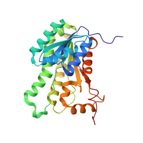
    S-(+)-1-Amino-2-propanol dehydrogenase (APDH) is a short-chain dehydrogenase/reductase associated with the incompletely characterized Rhodococcus and Mycobacterium bacterial microcompartment (RMM). We enzymatically characterized the APDH from M. smegmatis and showed it is highly selective, with a low micromolar K m for S-(+)-1-amino-2-propanol and specificity for NADP(H). A paralogous enzyme from a nonmicrocompartment-associated operon in the same organism was also shown to have a similar activity. We determined the structure of APDH in both apo form (at 1.7 Å) and as a ternary enzyme complex with NADP + and aminoacetone (at 1.9 Å). Recognition of aminoacetone was mediated by strong hydrogen bonds to the amino group by Thr145 and by Glu251 from the C-terminus of an adjacent protomer. The substrate binding site entirely encloses the substrate, with close contacts between the aminoacetone methyl group and Phe95, Trp154, and Leu195. Kinetic characterization of several of these residues confirm their importance in enzyme functioning. Bioinformatics analysis of APDH homologues implies that many nonmicrocompartment APDH orthologues partake in an aminoacetone degradation pathway that proceeds via an aminopropanol O-phosphate phospholyase. RMM microcompartments may mediate a similar pathway, though possibly with differences in the details of the pathway that necessitates encapsulation behind a shell.


  • Organizational Affiliation

    Department of Molecular and Cellular Biology , University of Guelph , Guelph , Ontario N1G 2W1 , Canada.


Macromolecules
Find similar proteins by:  (by identity cutoff)  |  3D Structure
Entity ID: 1
MoleculeChains Sequence LengthOrganismDetailsImage
3-oxoacyl-[acyl-carrier-protein] reductase
A, B, C, D, E
A, B, C, D, E, F, G, H, I, J, K, L, M, N, O, P
259Mycolicibacterium smegmatis MC2 155Mutation(s): 0 
Gene Names: MSMEG_0269
EC: 1.1.1.100
UniProt
Find proteins for A0QP46 (Mycolicibacterium smegmatis (strain ATCC 700084 / mc(2)155))
Explore A0QP46 
Go to UniProtKB:  A0QP46
Entity Groups  
Sequence Clusters30% Identity50% Identity70% Identity90% Identity95% Identity100% Identity
UniProt GroupA0QP46
Sequence Annotations
Expand
  • Reference Sequence
Small Molecules
Ligands 4 Unique
IDChains Name / Formula / InChI Key2D Diagram3D Interactions
NAP (Subject of Investigation/LOI)
Query on NAP

Download Ideal Coordinates CCD File 
AB [auth K]
CA [auth D]
DB [auth L]
FA [auth E]
HB [auth M]
AB [auth K],
CA [auth D],
DB [auth L],
FA [auth E],
HB [auth M],
IA [auth F],
LB [auth N],
MA [auth G],
OB [auth O],
PA [auth H],
Q [auth A],
SB [auth P],
TA [auth I],
V [auth B],
WA [auth J],
Y [auth C]
NADP NICOTINAMIDE-ADENINE-DINUCLEOTIDE PHOSPHATE
C21 H28 N7 O17 P3
XJLXINKUBYWONI-NNYOXOHSSA-N
F3V (Subject of Investigation/LOI)
Query on F3V

Download Ideal Coordinates CCD File 
BB [auth K]
DA [auth D]
EB [auth L]
GA [auth E]
IB [auth M]
BB [auth K],
DA [auth D],
EB [auth L],
GA [auth E],
IB [auth M],
JA [auth F],
MB [auth N],
NA [auth G],
PB [auth O],
QA [auth H],
R [auth A],
TB [auth P],
UA [auth I],
W [auth B],
XA [auth J],
Z [auth C]
1-aminopropan-2-one
C3 H7 N O
BCDGQXUMWHRQCB-UHFFFAOYSA-N
CL
Query on CL

Download Ideal Coordinates CCD File 
AA [auth C]
CB [auth K]
EA [auth D]
FB [auth L]
HA [auth E]
AA [auth C],
CB [auth K],
EA [auth D],
FB [auth L],
HA [auth E],
JB [auth M],
KA [auth F],
NB [auth N],
OA [auth G],
QB [auth O],
RA [auth H],
S [auth A],
T [auth A],
UB [auth P],
VA [auth I],
X [auth B],
YA [auth J]
CHLORIDE ION
Cl
VEXZGXHMUGYJMC-UHFFFAOYSA-M
MG
Query on MG

Download Ideal Coordinates CCD File 
BA [auth C]
GB [auth L]
KB [auth M]
LA [auth F]
RB [auth O]
BA [auth C],
GB [auth L],
KB [auth M],
LA [auth F],
RB [auth O],
SA [auth H],
U [auth A],
ZA [auth J]
MAGNESIUM ION
Mg
JLVVSXFLKOJNIY-UHFFFAOYSA-N
Experimental Data & Validation

Experimental Data

  • Method: X-RAY DIFFRACTION
  • Resolution: 1.90 Å
  • R-Value Free: 0.233 
  • R-Value Work: 0.204 
  • R-Value Observed: 0.205 
  • Space Group: P 1 21 1
Unit Cell:
Length ( Å )Angle ( ˚ )
a = 131.77α = 90
b = 129.06β = 107.88
c = 145.79γ = 90
Software Package:
Software NamePurpose
PHENIXrefinement
XDSdata reduction
XSCALEdata scaling
PHASERphasing
RESOLVEphasing
PDB_EXTRACTdata extraction

Structure Validation

View Full Validation Report


Ligand Structure Quality Assessment 


Entry History & Funding Information

Deposition Data


Funding OrganizationLocationGrant Number
Natural Sciences and Engineering Research Council (NSERC, Canada)Canada04045-2015

Revision History  (Full details and data files)

  • Version 1.0: 2018-05-30
    Type: Initial release
  • Version 1.1: 2018-07-11
    Changes: Data collection, Database references
  • Version 1.2: 2019-02-20
    Changes: Author supporting evidence, Data collection
  • Version 1.3: 2020-01-08
    Changes: Author supporting evidence
  • Version 1.4: 2023-10-04
    Changes: Data collection, Database references, Derived calculations, Refinement description